Cellular Division
Every human cell plays a vital role in sustaining the body, with specialized types such as nerve, blood, and muscle cells. Each cell absorbs nutrients through its membrane, critical for energy production and molecular synthesis, which is necessary for survival. Protein synthesis, regulated by DNA in the nucleus, is essential for cell function. This article details the phases of cell division, including mitosis and meiosis, and explores the importance of each phase in maintaining healthy cellular function and the implications of radiation on cellular integrity.

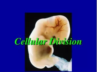
Cellular Division
E N D
Presentation Transcript
Cell Function Every human cell has a specific function in supporting the total body. Some differences are obvious, as in nerve cells, blood cells, and muscle cells. Similarities are also somewhat obvious In addition to its specialized function, each cell to some extent absorbs all molecular nutrients through the cell membrane and uses these nutrients in energy production and molecular synthesis. If this molecular synthesis is damaged by radiation exposure, the cell may malfunction and die.
Protein synthesis is a good example of a critical cellular function necessary for survival. DNA, located in the nucleus, contains a molecular code that identifies which proteins the cell will make. This code is determined by the sequence of base pairs (adenine–thymine and cytosine–guanine). A series of three base pairs, called a codon, identifies one of the 22 human amino acids available for protein synthesis. This genetic message is transferred within the nucleus to a molecule of mRNA. mRNA leaves the nucleus by way of the endoplasmic reticulum and makes its way to a ribosome, where the genetic message is transferred to yet another RNA molecule (tRNA). tRNA searches the cytoplasm for the amino acids for which it is coded. It attaches to the amino acid and carries it to the ribosome, where it is joined with other amino acids in sequence by peptide bonds to form the required protein molecule. Interference with any phase of this procedure for protein synthesis could result in damage to the cell. Radiation interaction in which the molecule has primary control over protein synthesis (DNA) is more effective in producing a response than is radiation interaction with other molecules involved in protein synthesis
Cell division is the multiplication process whereby one cell divides to form two or more cells. Mitosis (M) and meiosis are the two types of cell division that occur in the body. When somatic cells (all cells in the human body except the germ cells) divide, they undergo mitosis. Genetic cells (the oogonium, or female germ cell, and the spermatogonium, or male germ cell) undergo meiosis.
Although many thousands of rad (many gray) are necessary to produce physically measurable disruption of macromolecules in vitro, single ionizing events at a particularly sensitive site of a critical target molecule are thought to be capable of disrupting cell proliferation.
Mitosis Through the process of mitosis (M), a parent cell divides to form two daughter cells identical to the parent cell. This process results in an approximately equal distribution of all cellular material between the two daughter cells. The cellular life cycle may be pictured as in. Different phases of cell growth, maturation, and division occur in each cell cycle. Four distinct phases of the cellular life cycle are identifiable: M (mitosis phase), G1(pre-DNA synthesis phase), S (synthesis phase), and G2 (post-DNA synthesis phase). Additionally, mitosis (M) can be divided into four subphases: prophase, metaphase, anaphase, and telophase.
Mitosis is the division phase of the cellular life cycle. It is actually the last phase of the cycle. After it has commenced, it takes only about 1 hour to complete in all cells. Interphase, the period of cell growth that occurs before actual mitosis, consists of three intervals: G1, S, and G2. G1, the earliest, is the phase between reproductive events. It is the gap in the growth of the cell that occurs between mitosis and DNA synthesis. Depending on the type of cells involved, this interval may take just a few minutes or it may take several hours. G1 is designated as the pre-DNA-synthesis phase. During G1 a form of RNA is synthesized in the cells that are to reproduce. This RNA is needed before actual DNA synthesis can efficiently begin.
The cell biologist usually identifies four phases of the cell cycle: M, G1, S, and G2. These phases of the cell cycle are characterized by the structure of the chromosomes, which contain the genetic material DNA. The gap in cell growth between M and S is G1. G1 is the pre-DNA synthesis phase. The DNA synthesis phase is S. During this period, each DNA molecule is replicated into two identical daughter DNA molecules During S phase, the chromosome is transformed from a structure with two chromatids attached to a centromere to a structure with four chromatids attached to a centromere.The result is two pairs of homologous chromatids, that is, chromatids with precisely the same DNA content and structure. The G2 phase is the post-DNA synthesis gap of cell growth During interphase, the chromosomes are not visible; however, during mitosis, the DNA slowly takes the form of the chromosomes as seen microscopicallyschematically depicts the process of mitosis. .
While the S phase is taking place, the chromosome changes in shape from a figure with two chromatids connected to a centromere to a figure with four chromatids connected to a centromere.
Prophase During prophase, the first phase of cell division, the nucleus enlarges, the DNA complex (the chromatid network of threads) coils up more tightly, and the chromatids become more visible on stained microscopic slides. Chromosomes enlarge, and the DNA begins to take structural form. The nuclear membrane disappears, and the centrioles (small hollow cylindrical structures) migrate to opposite sides of the cell and begin to regulate the formation of the mitotic spindle, the delicate fibers that are attached to the centrioles and extend from one side of the cell to the other across the equator of the cell.
Metaphase As metaphase begins, the fibers collectively referred to as the mitotic spindle form between the centrioles. Each chromosome (which now consists of two chromatids) lines up in the center or equator of the cell attached by its centromere to the mitotic spindle. This forms the equatorial plate. The centromeres then duplicate, and each chromatid attaches itself individually to the spindle. At the end of metaphase, the chromatids are strung out along the mitotic spindle much like laundry hung on a clothesline. During metaphase, cell division can be stopped and visible chromosomes can be examined under a microscope. Chromosome damage caused by radiation can then be evaluated.
Anaphase During anaphase, the duplicate centromeres migrate in opposite directions along the mitotic spindle, carrying the chromatids to opposite sides of the cell. The cell is now ready to begin the last phase of division.
Telophase During telophase, the chromatids undergo changes in appearance by uncoiling and becoming long, loosely spiraled threads. Simultaneously the nuclear membrane re-forms, and two nuclei (one for each new daughter cell) appear. The cytoplasm also divides (cytokinesis) near the equator of the cell to surround each new nucleus. After this cell division completes, each daughter cell has a complete cell membrane and contains exactly the same amount of genetic material (46 chromosomes) as the parent cell
Sensitivity – Cell Cycle Phase • Cells are most sensitive to radiation during mitosis (M phase) and RNA synthesis (G2 phase) • Less sensitive during the preparatory period for DNA synthesis (G1 phase) • Least sensitive during DNA synthesis (S phase) • During mitosis (M), the metaphase is the most sensitive
Radiation-induced chromosome damage is analyzed during metaphase.
Meiosis Meiosis is a special type of cell division that reduces the number of chromosomes in each daughter cell to half the number of chromosomes in the parent cell. Male and female germ cells, or sperm and ova, of sexually mature individuals each begin meiosis with 46 chromosomes. However, before the male and female germ cells unite to produce a new organism, the number of chromosomes in each must be reduced by one half to ensure that the daughter cells (zygotes) formed when they unite will contain only the normal number of 46 chromosomes. Hence, meiosis is really a process of reduction division. .
Paradoxically, meiosis begins with a doubling of the amount of genetic material; as in mitosis, DNA replication occurs during interphase. As a result of DNA replication, each one-chromatid chromosome duplicates, forming a two-chromatid chromosome. This means that sperm and egg cells begin meiosis with twice the amount of genetic material as the original parent cell. Thus, at the beginning of meiosis, the number of chromosomes increases from 2n to 4n (n = 23). The various phases of meiosis are similar to those occurring in mitosis. The major difference between the two types of cell division begins at the end of telophase. In meiosis, after the parent germ cell has formed two daughter cells, each of which (in human beings) contains 46 chromosomes, the daughter cells divide without DNA replication; chromosome duplication does not occur at this phase of division. These two successive divisions result in the formation of four granddaughter cells, each of which contains only 23 chromosomes. This means that the proper number of 46 chromosomes will be produced when a female ovum containing 23 chromosomes is fertilized by a male sperm containing 23 chromosomes
TISSUES AND ORGANS During the development and maturation of a human from two united genetic cells, a number of different types of cells evolve. Collections of cells of similar structure and function form tissues. These tissues in turn are precisely bound together to form organs. The tissues and the organs of the body serve as discrete units with specific functional responsibilities. Some tissues and organs combine into an overall integrated organization known as an organ system. The principal organ systems of the body are the nervous system, the digestive system, the endocrine system, the respiratory system, and the reproductive system. Effects of radiation that appear at the whole-body level result from damage to these organ systems that occurs as the result of radiation injury to the cells of that system.
The cells of a tissue system are identified by their rate of proliferation and their stage of development. Immature cells are called undifferentiated cells, precursor cells, or stem cells. As a cell matures through growth and proliferation, it can pass through various stages of differentiation into a fully functional and mature cell Stem cells are more sensitive to radiation than mature cells
In 1906, two French scientists, BergonieandTribondeau, theorized and observed that radiosensitivity was a function of the metabolic state of the tissue being irradiated. This has come to be known as the Law of Bergonieand Tribondeau and has been verified many times. Basically, the law states that: • the radiosensitivity of cell is directly proportional to their reproductive activity and inversely proportional to their degree of differentiation. • Cells most active in reproducing themselves and cells not fully mature will be most harmed by radiation. • The more mature and specialized in performing functions as cell is, the less sensitive it is to radiation. • This law is principally interesting as a historical note in the development of radiobiology. It has found some application in radiation oncology. In diagnostic imaging, the law serves to remind us that the fetus is considerably more sensitive to radiation exposure than the child or the mature adult
Radiosensitivity varies with age. Experiments with animals have shown that the very young and the very old are more sensitive to radiation
The sensitivity of the cell to radiation is determined somewhat by its state of maturity and its functional role. The tissues and organs of the body include both stem cells and mature cells. Several types of tissue can be classified according to structural or functional features. These features influence the degree of radiosensitivity of the tissue.