Download
1 / 1
10 likes | 172 Vues
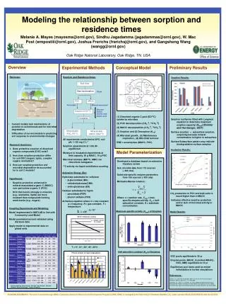
84,000cfu. 9,700cfu. 3,000cfu. 1,360cfu. 220cfu. Sample 1. Sample 2. RLU. Control. Time (sec). III. Reproducibility of the CANARY TM biosensor response

E N D
84,000cfu 9,700cfu 3,000cfu 1,360cfu 220cfu Sample 1 Sample 2 RLU Control Time (sec) III. Reproducibility of the CANARYTM biosensor response Various concentrations of E. coli O157:H7 were added to assay buffer and centrifuged at 10,000 x g to pellet the bacteria. Biosensor cells specific for E. coli O157:H7 were added and the sample was centrifuged for an additional 5 seconds, then placed in the luminometer to record the response. CFU E. coli RLU 5000 500 50 0 Time (sec) Application of a biosensor for rapid detection of E. coli O157:H7contamination in foodKevin J. Modarress, Iwona Mielzynska, Qiao-xi Zheng, and Thomas HazelInnovative Biosensors, Inc., College Park, MD Introduction Development of rapid, sensitive methods for detecting E. coli O157:H7 is of significant interest because it offers the opportunity to increase the efficiency of food testing and decrease risk to the public. We offer a novel biosensor system that allows for simple, sensitive, real-time detection of pathogens in a variety of food matrixes. The sensor, based on the CANARYTM technology, allows for the detection of as few as 50cfu E. coli O157:H7 and requires only 5 minutes to perform. Our objective was to characterize the sensitivity of the CANARYTM assay in detecting E. coli O157:H7 in ground beef samples. The CANARYTM technology consists of a cell line that is genetically engineered to recognize a specific pathogen, responding to its presence by emitting a luminescent signal that can be detected using a standard luminometer. The potential of this technology has been demonstrated by its application to the detection of 20 different viral and bacterial pathogens to date (Rider et al., 2003). In both simple and complex samples CANARYTM biosensor cells demonstrate sensitivity and specificity that rivals PCR in the detection of such pathogens as E. coli O157:H7 and Bacillus anthracis. Furthermore, this technology provides results in as little as 2 minutes and can be applied by individuals with a minimum of technical expertise, making it ideal for routine application in many settings. Construction of new biosensor cell lines is relatively simple and requires only that a monoclonal antibody be available for the target of interest. cDNAs encoding the light and heavy chains of the antibody are cloned into a vector that targets antibody to the cell surface. These vectors are transfected into a parental cell line expressing a bioluminescent protein, wherein antibodies are expressed and localized to the cell surface. Exposure of the biosensor cell line to its target pathogen triggers release of calcium from internal stores, thus activating the luminescent properties of the marker protein (see figure below). IV. Sensitivity of the CANARYTME. coli O157:H7 biosensor Various concentrations of E. coli O157:H7 were added to assay buffer and analyzed with CANARYTM biosensor as described in Experimental Methods. Results are expressed as signal/noise (S/N), a ratio of the luminescent signal (in RLU’s) in the presence of bacteria to that in the presence of assay buffer alone. VII. Biosensor detection of E. coli O157:H7 in ground beef Ground beef samples were spiked with 0.8cfu/g of E. coli O157:H7, followed by 6 hours enrichment as described in Experimental Methods. The samples were then processed for detection with the CANARYTM biosensor. Each sample was tested in triplicate and compared to an unspiked negative control. Results are expressed as Relative Light Units (RLU). I. Experimental Methods Measurement of pathogen in non-complex samples. Enumerated E. coli were spiked in 250 ml assay buffer at various concentrations and samples were centrifuged for 2 minutes at 10,000 x g. Biosensor reagent was added to the tube containing the sample for testing and the sample was centrifuged for 5 seconds, then placed in a single-tube luminometer and luminescence measured for a total of 60 seconds. Measurement of pathogen in ground beef. Ground beef samples obtained from a commercial outlet were placed in sterile stomacher filter sample bags (25g of ground beef per bag) and inoculated with E. coli O157:H7. Samples were diluted in enrichment medium (225mL of mEC without novobiocin), agitated using a Stomacher device and incubated for various time at 37ºC. After enrichment 3ml of sample was removed from the clean compartment of the filter bag, placed in a sterile tube and left undisturbed for 2-4 minutes to allow particulate material to settle. 1ml of sample was then transferred into 1.5ml centrifuge tubes and spun down at 10,000 x gfor 2minutes, then washed twice with PBS+0.05% Tween-20 and centrifuged again. The supernatant was removed and replaced with 250mL of assay buffer and samples were centrifuged again at 10,000 x g for 2 minutes, then assayed using CANARYTM biosensor cells. Ground beef samples (25g) inoculated with E. coli O157:H7 (0.8 cfu/g) were assayed using CANARYTM after various times of enrichment and the number of viable E. coli O157:H7 cells determined empirically. Results are expressed as a ratio of signal to noise for a given data point, with a value of 3 (dashed line) considered positive. Values shown are the average of at least 3 measurements. V. Specificity of the CANARYTME. coli O157:H7 biosensor Randomly selected non-O157 E. coli isolates were tested using the CANARYTME. coli O157:H7 biosensor at densities of 107cfu per reaction. The biosensor shows virtually no cross-reactivity with these strains. II. Procedure for detection of E. coli O157:H7 using CANARYTM CANARYTM provides a rapid method for detecting the presence of E. coli O157:H7 in complex and non-complex samples. In non-complex matrixes the sample is centrifuged at 10,000 x g to pellet the pathogen. Biosensor is added and the sample centrifuged briefly to bring the biosensor in contact with the pathogen, initiating the luminescent response. Pathogen Non-complex sample S/N Total assay time ~3 minutes Add 1 drop of sample Add 1 drop of Biosensor Centrifuge sample Centrifuge sample IP3 Detect luminescence 2 min 5 sec Ca2+ CFU E. coli O157:H7 Bioluminescence Conclusions Principal advantages of the CANARYTM technique include its speed and sensitivity. Even in complex samples such as ground beef, this technology enables rapid detection of pathogens. The results presented here demonstrate that CANARYTM has the capacity to detect as few as 50cfu E. coli O157:H7 in non-complex samples and 220cfu E. coli O157:H7 in ground beef samples in 5 minutes or less with a minimum of sample processing. The CANARYTM assay shows sensitivity that is comparable to that of PCR (Cui et al., 2003; Ellingson et al., 2005; Li and Drake, 2003), with the capacity to detect low levels of E. coli O157:H7 contamination (<1cfu/g) in ground beef samples after an enrichment time of 7hr or less. The speed and simplicity with which CANARYTM can be performed is a significant advantage over the laborious and time-consuming steps that are required for PCR. Schematic of the CANARYTM Technology VI. Rapid processing of samples for detection of E. coli O157:H7 in ground beef After enrichment (see Experimental Methods) 1ml of the sample is pelleted by centrifugation at 10,000 x g. The supernatant is then discarded and the sample washed twice with wash buffer. The sample is centrifuged again and the wash buffer replaced with a pH-neutral assay buffer. Biosensor is added and a brief centrifugation step brings the biosensor in contact with the sample, initiating the luminescent response. BioFlashTM Instrumentation Equipment required for detection of the CANARYTM response is off-the-shelf technology. A high-speed benchtop microfuge is used to concentrate pathogen at the bottom of a tube and the biosensor cells are brought into contact with the pathogen using a small minifuge with a horizontal rotor. A small single-tube luminometer records the response of the biosensor cells, and data is stored and analyzed on a laptop computer. References Cui S., C.M. Schroeder, D.Y. Zhang, and J. Meng. 2003. Rapid sample preparation method for PCR-based detection of Escherichia coli O157:H7 in ground beef. J Appl Microbiol. 95:129-134. Ellingson J.L., J.J. Koziczkowski, J.L. Anderson, S.A. Carlson, and V.K. Sharma. 2005. Rapid PCR detection of enterohemorrhagic Escherichia coli (EHEC) in bovine food products and feces. Mol. Cell. Probes 19:213-217. Li W., and M.A. Drake. 2003. Detection of viable Shiga toxin-producing Escherichia coli by quantitative competitive polymerase chain reaction. J Food Prot. 66:1277-1282. Rider, T.H., M.S. Petrovick, F.E. Nargi, J.D. Harper, E.D. Schwoebel, R.H. Mathews, D.J. Blanchard, L.T. Bortolin, A.M. Young, J. Chen, and M.A. Hollis. 2003. A B cell-based sensor for rapid identification of pathogens. Science 301:213-215. Complex sample Total assay time ~5 minutes add 1 drop of Biosensor Add 1mL of sample Remove liquid & wash Centrifuge sample Centrifuge sample centrifuge sample Detect luminescence 2 min 5 sec Replace buffer Contact Information: Thomas Hazel tom.hazel@innovativebiosensors.com www.innovativebiosensors.com Instrumentation required for use with CANARYTM Technology