Intraoral
Intraoral. Intraoral imaging briefly. Dental Radiography Radiographs enable the dental professional to identify many conditions that cannot be identified through visual inspection. Radiographs allow the inspection of the tooth structure and supporting bone. Intraoral imaging briefly.

Intraoral
E N D
Presentation Transcript
Intraoral imaging briefly Dental Radiography • Radiographs enable the dental professional to identify many conditions that cannot be identified through visual inspection. • Radiographs allow the inspection of the tooth structure and supporting bone.
Intraoral imaging briefly Assessment and Detection of: • Missing teeth • Unerupted teeth • Impacted teeth • Dental caries • Periodontal disease • Tooth abnormalities • Retained roots • Cysts and tumors
Selling Digital There are four aspects to position digital radiography… Convenience Financial/business Better Medicine The Environment
Selling Digital Convenience • Instant diagnosis (versus film and scanners) • Instant error correction (retaking is very easy) • No film • No ongoing and unpredictable costs • No chemicals (storage, handling, processing, disposal) • No walking back and forth to darkroom…and waiting…and waiting…and waiting • On a monitor • Easier to see any issues and/or pathology • Image manipulation
Selling Digital Better medicine • Easier to communicate with patient • Higher patient case acceptance • Significantly lower radiation than film • Tools that are not available with film…
Selling Digital Full screen images make it easy to demonstrate diagnosis to patient If the patient can see the problem, they are more likely to accept treatment.
Selling Digital Demonstrate the issue to the patient Measure working length Ensure the apex has been reached Review and present to the patient
Selling Digital The environment • No chemicals! • No wasteful consumables
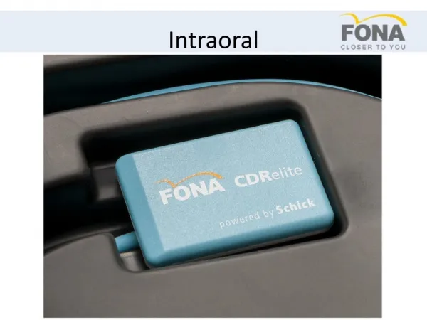
Introduction to Products FONA CDR and FONA CDRelite powered by Schick
Products FONA CDRelite • Imaging system launched in 2009 • 2nd generation CMOS-APS technology • Three film like sizes to suit all patient sizes (0, 1 and 2) • Connection to the computer via a remote requiring power ONLY from USB port (USB 1.1 and 2.0) • Theoretical resolution of 16.7 lp/mm and actual resolution of 16.0 lp/mm • Pixel size of 30μm • Durable plastic sensor can (not susceptible to bite marks, scratches, nicks) • Unique replaceable cable (new sensor kits include a spare cable) • Modular platform • Gadox scintillator • Edge enhancement options to suit clinician’s personal preferences • Premium, customizable solution.
FONA CDRelite Connection location minimizes cable interference during positioning (ideal for vertical bitewings) Bright colors for increased visibility in the oral cavity Curved corners for maximum patient comfort
FONA CDRelite Replaceable cable
Selling FONA CDR • Best image at price point • Available in three sizes to fit all patient types • Also allows for versatility in selling just one sensor as needed (i.e. in India being price-sensitive we essentially sell just size 1 sensors) • Modularity of system • Remote module is separate from the sensor. As technology changes, customers only need to upgrade the component that becomes obsolete*
Selling FONA CDRelite Superior image quality Cavity Detection Bone Tribeculation Lamina Dura and Apex Detail
Unenhanced Selling FONA CDRelite
Edge Smooth(Noise free, film-like appearance) Selling FONA CDRelite
Edge low(Edge definition with low noise) Selling FONA CDRelite
Edge high(maximum edge definition with noise) Selling FONA CDRelite
Selling FONA CDRelite Direct USB Connection • USB connectors are only required to last 1500 insertion/extraction cycles • If the USB connector fails, it requires the replacement of the entire sensor. • Schick’s HypertronicsHypertac connectors are proofed to 20,000 cycles. • Gold-plated connectors • Low insertion force • Long contact life • Should a failure occur to the connector, the cable can instantly be replaced.
Selling FONA CDRelite • Kink resistant outer jacket • Thick jacket for greater protection • Cable Replacement • Less than one minute • Every sensor shipped with a spare replaceable cable. • Cables available in 3 lengths for maximum convenience 3’ / 0.9m 6’ / 1.8m 9’ / 2.7m
Vs The Competition * Claimed by manufacturer without proof
Summary – Why Digital? • Return on Investment • Predictable expense • Increased workflow • Increased patient acceptance • Increased revenue • Instant imaging and retakes • No film or chemicals • Enhanced diagnostics • Simple referrals • Lower radiation
Summary – Why FONA CDRelite? • Competitive premium imaging at lower prices than competition • 3 sizes to fit every patient • 3 cable lengths for the needs of the practice • Spare cable with each new sensor • Risk mitigation with one step cable replacement • Design focused on easy positioning • Hypertac connectors rated far more durable • Modular platform so that components that break are all that need to be replaced…not the entire system
Positioning Schick designed parallel-technique based holder system, available in one of two forms… Adhesive Unique, patented design based on the RINN system, adaptable to suit different anatomies; maximizes patient comfort. Grip Completely new design offers a robust, easy to assemble, autoclavable holder solution for FONA CDRelite sensors
Positioning • Three key things to remember… • Become a positioning expert. The better you are, the easier to sell • NEVER, EVER, EVER SAY “BITE”…always ask the patient to “close to pressure” • Always use a barrier / sheath
Positioning Maxillary Anterior Placement Place the distal end of the sensor against the roof of the mouth, with the incisal edge of the teeth against the front of the tab. Sensor should be parallel to the long axis of the maxillary anterior teeth. Ensure the ring is as close to the patient’s face as possible and place the x-ray head against the ring.
Positioning Mandibular Anterior Placement Place the sensor into the lower anterior area, positioning it on top of the tongue, parallel to the first molar. Sensor should be centered on the mandibular anterior teeth when the patient is occluded. Ensure the ring is as close to the patient’s face as possible and place the x-ray head against the ring.
Positioning Horizontal Bitewing Placement To capture a horizontal bitewing image, place the sensor between the tongue and the teeth with the bite area resting on the premolar teeth. The patient should close on their back teeth to ensure centric occlusion and as they do so, the arm should be angled gently toward the midline of the mouth to ensure the sensor is parallel with the teeth and to provide open contacts. Ensure the ring is as close to the patient’s face as possible and place the x-ray head against the ring.
Positioning Vertical Bitewing Placement The sensor should enter the mouth horizontally. Once past the incisors, “roll” it into a vertical position. The sensor should be placed with the cable pointing upwards toward the hard palate. Ensure the ring is as close to the patient’s face as possible and place the x-ray head against the ring.
Positioning Maxillary Posterior Placement The sensor/aiming device is angled upward toward the midline with placement of the bite block under the teeth to be captured. The sensor should be angled slightly past the midline of the palate as the patient closes for comfort and to ensure capture of the apices. Ensure the ring is as close to the patient’s face as possible and place the x-ray head against the ring.
Positioning Mandibular Periapical Placement Retract the cheek with a finger and place the sensor between the tongue and the teeth, bringing the cheek around the bite block. Then slide the sensor down and in gently until it is in position—the bite tab should be directly above the teeth to be imagined. Ensure the ring is as close to the patient’s face as possible and place the x-ray head against the ring.
Positioning • AGAIN, three key things to remember… • Become a positioning expert. The better you are, the easier to sell • NEVER, EVER, EVER SAY “BITE”…always ask the patient to “close to pressure” • Always use a barrier / sheath