Download
1 / 1
30 likes | 176 Vues
Increased Expression of Secreted Frizzled-Related Protein-1 and Microtubule-Associated Protein Light Chain 3 in Keratoconus Omer Iqbal, MD, George Fisher, MD, Samir Vira, MD, Daneyal Syed, BS, Nasir Sadeghi, MS, David Freeman, BS, Edward Campbell, PhD, Joel Sugar MD,

E N D
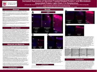
Increased Expression of Secreted Frizzled-Related Protein-1 and Microtubule-Associated Protein Light Chain 3 in Keratoconus Omer Iqbal, MD, George Fisher, MD, Samir Vira, MD, Daneyal Syed, BS, Nasir Sadeghi, MS, David Freeman, BS, Edward Campbell, PhD, Joel Sugar MD, Robert Feder, MD, Jawed Fareed, Ph.D, Charles Bouchard, MD Department of Ophthalmology/Pathology, Loyola University Health System, Maywood, IL. 60153. Results Abstract LC3 Purpose: To study the expression of secreted frizzled related protein-1 (SFRP-1) and microtubule-associated protein light chain-3 (LC3), an autophagy marker, in keratoconus. Methods: Under an IRB approved protocol, de-identified and/or surgically discarded normal donor (n=10) and keratoconus corneas (n=10) were obtained. The corneal samples were fixed in formalin and embedded in paraffin. Immunohistochemical staining using SFRP-1 and LC3 antibodies was performed. Results: The majority of expression of SFRP1 was seen in the epithelium; however, in 3 tissues that showed high expression, staining was also present in the stroma and endothelium. Like SFRP1, the LC3 expression in keratoconus tissues occurred at 3 different levels: low, medium, and high. Collectively these data suggest that there are differences in expression levels of SFRP1 and LC3 in keratoconus tissue compared to normal tissue. Low expressivity of SFRP-1 appeared to correspond with low expressivity of LC3 while medium-high expressivity of SFRP-1 corresponded with medium to high expressivity of LC3. Conclusions: Increased expression of SFRP-1 and LC3 was observed in keratoconus corneas. Keratocyte autophagy seen with keratoconus may play a role in the pathogenesis of keratoconus. Keywords: Keratoconus; Keratocytes; Apoptosis; Autophagy; Immunohistochemistry. SFRP Introduction Keratoconus is a non-inflammatory, bilateral, progressive, often asymmetric, primary ectasia associated with irregular astigmatism and decreased visual acuity. It is one of the most common indications for corneal transplantation. First described by Burchard Mauchart in 1748, keratoconus affects 1 in every 2000 Americans (1,2). Despite significant research, the pathogenesis of keratoconus is still poorly understood. Earlier studies suggest a role of oxidative stress and keratocyte apoptosis in the pathogenesis of keratoconus (13-16). A recent investigation of the expression of Wnt signaling pathway genes in keratoconic epithelium has been reported (17). In this study, the secreted frizzled related protein-1 (SFRP-1) was reported to be upregulated by 25-fold in the corneal epithelium of keratoconus patients (17). Based on this background it is hypothesized that dysregulation of Wnt signaling through aberrant up or down regulation of SFRP-1 may contribute to the pathogenesis of keratoconus. The aim of our study is to determine the SFRP-1 expression by immunohistochemical staining of formalin-fixed, paraffin embedded slides of normal, keratoconic and diseased corneas such as Fuchs’ dystrophy, lattice dystrophy and corneal edema. Furthermore, since autophagy has a significant association with programmed cell death or apoptosis (33), the role of microtubule-associated protein light chain 3 (LC3-II), an autophagosomal marker, will be explored in the pathogenesis of keratoconus. Autophagy entails bulk degradation of proteins and organelles which is crucial for cellular maintenance, cell viability, differentiation and development (34). Figure 2: A. Keratoconus tissues tagged with the LC3 antibody displayed from left to right in varying degrees of LC3 expression (pink) with the epithelium (Ep) on the top and correlating stroma (S) and endothelium (En) on the bottom. Note that the first two tissues displayed did not have identifiable endothelium. Low expression (tissue K9) displayed on the left, medium expression (tissue K3) displayed in the middle and high expression (tissue K8) displayed on the right. B. Normal tissues tagged with the LC3 antibody displayed from left to right in varying degrees of expression with the epithelium on the top and correlating stroma on bottom. These samples did not have identifiable endothelium. Low expression (tissue N1) displayed on the left, medium expression (tissue N3) displayed in the middle and no tissues of high expression. Cell nuclei were stained with DAPI in blue. Materials and Methods Corneal Samples Acquisition: A total of 12 normal donor corneas and 13 keratoconic corneas were obtained for the study. In addition, other diseased corneas, including 5 with Fuchs’ dystrophy, 4 with corneal edema, 1 each with lattice dystrophy, corneal amyloidosis and bullous keratopathy were also collected. The samples were immediately fixed in formalin and kept refrigerated until all the samples were collected. The tissue samples were embedded in paraffin blocks and sectioned using the microtome and slides were made. Immunohistochemical staining using SFRP-1 antibody: Tissue sections were deparaffinized by three 5 minute washes in xylene, followed by two washes in 100% ethanol for 2 minutes each, then washed in 95% ethanol for 5 minutes, followed sequentially by 70% ethanol for 5 minutes, distilled water for 1 minute and phosphate buffered saline (PBS) for 5 minutes. Sections were blocked with normal goat serum for 1 hour and were then washed and incubated with SFRP-1 primary antibody for 30 minutes in a humidified chamber at 4°C. The slides were then washed 3 times in PBS and were incubated with a secondary antibody, biotinylated goat anti-rabbit IgG and diamino-2 phenylindole (DAPI). After washing, the slides were processed using Avidin/Biotinylated enzyme Complex (ABC) kits from Vector Laboratories (Burlingame, CA) as per the manufacturer’s instructions. Sample color was developed with 3, 3’-diaminobenzidine (DAB) reagent from the Vector Laboratory. Immunohistochemical staining using LC3 antibody: Tissue sections were blocked for 20 minutes in PBS with 10% normal donkey serum (NDS), 0.1%NaN3, and 0.1% Saponin. Next, sections were incubated with LC3 (1:400) primary antibody for 30 minutes. Samples were then washed and incubated for 20 min in Cy5-donkey anti-rabbit secondary antibody (1:400, Jackson ImmunoResearch, Westgrove PA) and diamino-2 phenylindole (DAPI). After washing in PBS, the slides were mounted using Fluoro-gel (Electron Microscopy Services, Hatfield PA). All images were obtained on a DeltaVision microscope. Within each experiment, image acquiring conditions were established and uniformly utilized to image all specimens in that experiment. Exposure times were kept consistent in processing all samples. Following deconvolution using the SoftWoRx deconvolution software (Applied Precision, Issaquah, WA), the corneas were analyzed in a systematic fashion that imaged the epithelium, stroma, and endothelium. Table 1. Summary of Immunostaining Results Figure 1. A. Keratoconus tissues tagged with the SFRP-1 antibody displayed from left to right in varying degrees of expression (pink) with the epithelium (Ep) on the top and correlating stroma (S) and endothelium (En) below. Note that the first two tissues displayed did not have endothelium. Low SFRP-1 expression (tissue K9) displayed on the left, medium expression (tissue K3) displayed in the middle and high expression (tissue K8) displayed on the right. B. Normal tissues tagged with the SFRP-1 antibody displayed from left to right in varying degrees of expression with the epithelium on the top and correlating stroma on the bottom. These samples did not have identifiable endothelium. Low expression (tissue N1) displayed on the left, medium expression (tissue N3) displayed in the middle and no tissues of high expression. C. Corneal edema tissue tagged with SFRP-1 antibody displayed from left to right with varying degrees of expression. These samples did not have identifiable endothelium. Low expression (tissue C4) displayed on the left, medium expression (tissue C2) displayed in the middle, and no tissues displayed high expression. Cell nuclei were stained with DAPI in blue. The table shows varying expression of SFRP-1 and LC3 in keratoconus, normal tissue, Fuchs dystrophy and corneal edema. Samples that showed autofluorescence are in bold. C, corneal edema; K, keratoconus; N, normal tissue. Conclusion The present study provided new information regarding the level of SFRP-1 and LC3 expression in the corneal epithelium and stroma of patient with keratoconus and adds to our understanding of the pathogenesis of this progressive disease process. Further investigations of the possible correlation of SFRP-1 and LC3 expression to the staging of the disease in the patients may allow us to develop a potential marker for disease progression. Additionally, SFRP-1 could serve as a potential target for the development of therapeutic drugs to halt the progression of keratoconus. Manipulation of SFRP-1 expression by Wnt pathway inhibitors or anti-β-catenin agents like micro-RNAs (MiR-2001, MiR-21 and DKK1) may offer a novel approach in the treatment of keratoconus (32). Similarly, significant attenuation of autophagy induced by starvation or rapamycin is reported to be achieved by protein kinase C activator, phorbo-12-myristate-13-acetate (PMA) or the protein phosphatase inhibitor, calyculin A (44). Key References 12. Sutton G, Madigan M, Roufas A et al. Secreted frizzled-related protein 1 (SFRP 1) is highly upregulated in keratoconus epithelium: a novel finding highlighting a new potential focus for keratoconus research and treatment. Clinical and Experimental Ophthalmology 2010;38(1):43-48. 17. Sugar J and Mascai MS. What Causes Keratoconus? Cornea 2012;31:716-719. 43. Kim WJ, Rabinowitz YS, Meisler DM and Wilson SE. Keratocyte apoptosis associated with Keratoconus. Exp. Eye Research 1999 69:475-481. All references are provided in the CORNEA Journal publication Acknowledgements: This work was supported by The Richard A. Perritt Charitable Foundation and Illinois Society for the Prevention of Blindness (ISPB).


