Download
1 / 25
250 likes | 354 Vues
CARTILAGE -SUPPORTING SPECIALIZED CONNECTIVE TISSUE -CONTAINS CHONDROBLASTS AND CHONDROCYTES -ABUNDANT EXTRACELLULAR MATRIX -HIGHLY HYDRATED -NOT VASCULARYZED. NOURISHED BY DIFFUSION. -A PERYCHONDRION IS PRESENT ALLOWING GROWTH IN YOUNG INDIVIDUALS

E N D
CARTILAGE -SUPPORTING SPECIALIZED CONNECTIVE TISSUE -CONTAINS CHONDROBLASTS AND CHONDROCYTES -ABUNDANT EXTRACELLULAR MATRIX -HIGHLY HYDRATED -NOT VASCULARYZED. NOURISHED BY DIFFUSION. -A PERYCHONDRION IS PRESENT ALLOWING GROWTH IN YOUNG INDIVIDUALS -A PERYCHONDRION IS NOT PRESENT IN ARTICULAR CARTILAGE
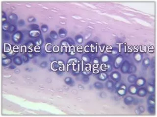
TYPES OF CARTILAGES HYALINE, (HIGH CONTENT IN COLLAGEN-NOT VISIBLE): MOST ABUNDANT. IT REPRESENTS THE FETAL SCHELETON. IT ALLOWS THE GROWTH IN LENGHT OF THE LONG BONES. IN THE ADULT IS PRESENT IN THE ARTICULATED JOINTS, IN TRACHEA, IN THE RIBS. ELASTIC, (HIGH CONTENT IN ELASTIC FIBERS-VISIBLE): EAR, EPYGLOTTIS. FIBROUS, (HIGH CONTENT IN COLLAGEN-VISIBLE): INTERVERTEBRAL DISCS, SYMPHYSIS, SOME TENDON-BONE ATTACHMENT. ALL THREE TYPES OF CARTILAGE PRESENT HIGH LEVELS OF PROTEOGLYCANS, EVENTHOUGH AT DIFFERENT CONCENTRATION.
HYALINE CARTILAGE A B Sections through hyaline cartilage. A, Low-power view of human rib, showing perichondrium (left), young chondroblasts embedded in pale-staining interterritorial matrix and mature chondrocytes embedded in the basophilic interterritorial matrix (centre and right). B, Higher magnification of hyaline cartilage in human bronchial wall, showing isogenous groups of chondrocytes. Note the more deeply-stained basophilic zones (rich in acidic proteoglycans) around the cell clusters, with older, paler-staining matrix between clusters.
CARTILAGE Elastic cartilage, stained to demonstrate elastin fibres (blueblack). Chondroblasts and larger chondrocytes are embedded in the matrix, which also contains collagen type II fibres. Highly cellular fetal cartilage, human phalanx. The cells are small and almost uniform in distribution. Mallory’s triple stain.
FIBROUS CARTILAGE White fibrocartilage in a late fetal intervertebral disc (human). Chondrocytes lie between coarse collagen type I fibres (blue) derived from the anulus fibrosus. Mallory’s triple stain.
CHONDROBLASTS Electron micrograph of chondroblasts in rabbit femoral condylar cartilage. The central cell has an active euchromatic nucleus with a prominent nucleolus, and its cytoplasm contains rough endoplasmic reticulum, scattered mitochondria, lysosomes and glycogen aggregates. The plasma membrane bears numerous short filopodia which project into the surrounding matrix. The latter shows a delicate feltwork of collagen fibrils within finely granular interfibrillary material. No pericellular lacuna is present; the matrix separates the central chondroblast from the cytoplasm of two adjacent chondroblasts (left, and crescentic profile).
BONE SUPPORTING AND PROTECTING CONNECTIVE TISSUE. MINERALYZED SOLID EXTRACELLULAR MATRIX Ca2+ AND PHOSPHATE ION DEPOSIT EXTRACELLULAR MATRIX CONTAINING 30% ORGANIC COMPONENTS AND 70% HYDROXYAPATITE CRISTALS CELLULAR COMPONENTS: 1 –PROGENITOR CELLS IN PERYOSTIUM AND ENDOSTIUM 2 -OSTEOBLASTS 3 -OSTEOCYTES 4 -OSTEOCLASTS
MORPHOLOGYCAL ELEMENTS OF BONE PERYOSTIUM: VASCULARYZED DENSE CONNECTIVE TISSUE CONTAINING PROGENITOR CELLS COVERING BONE ENDOSTIUM: THINNER, LAINING INTERNAL CAVITIES VOLKMAN AND HAVERS CHANNELS CONTAINIG BLOOD VESSELS NON LAMELLAR BONE (PHOETAL LIFE AND BONE REPAIR) LAMELLAR BONE (COMPACT AND SPONGIOSUM)
BONE REMODELLING BONE ABSORPTION PERFORMED BY OSTEOCLASTS UNDER CALCITONIN STIMULATION BONE DEPOSITION BY OSTEOBLAST SECRETION OF ORGANIC MATRIX AND MINERALYZATION TURNOVER OF MATRIX COMPONENTS BY OSTEOCYTES EACH YEAR 1/5 OF THE SCHELETON IS DEMOLISHED AND REBUILT
BONE FORMATION ENDOCHONDRAL OSSYFICATION: CHARTILAGE MOLD IS GRADUALLY SUBSTITUTED BY BONE DIRECT OXYFICATION: OSTEBLASTS DIFFERENTIATE IN A CONNECTIVE TISSUE MEMBRANE. (FLAT BONES) A NON LAMELLAR YOUNG BONE IS FIRST FORMED WHICH IS THEN SOBSTITUTED BY LAMELLAR BONE
BONE CAVITIES INTERNAL CAVITIES OF BONE CONTAIN BONE MARROW BONE MARROW CAN BE RED (HEMOPOYETIC TISSUE, ABUNDANT IN FLAT BONES) OR YELLOW (UNIVACUOLAR ADIPOCYTES, LONG AND SHORT BONES)
Vertical section 2 cm below the anterosuperior border of the iliac crest (female, 42 years). The cancellous bone consists of intersecting curved plates and struts. Osteonal (Haversian) canals can just be seen in the two cortices at this magnifi cation.
OSTEOCYTE LACUNAE Osteocyte lacunae shown at high magnification in a dry ground section of lamellar bone. The territories of three osteocytes are shown. Their branching dendrites contact those of neighbouring cells via the canaliculi seen here within the bone matrix. Several other osteocyte lacunae are present, out of the focal plane in this section, and tangential to the osteon axis.
NON LAMELLAR > LAMELLAR BONE Human parietal bone (male neonate) showing primary osteonal bone (grey) and woven (immature) bone (white) containing many connecting osteocyte lacunae (black). Internal resorption of the bone has produced large irregular dark spaces (trabecularization).
OSTEONES A, Osteons in a dry ground transverse section of bone. Concentric lamellae surround the central Haversian canal of each complete osteon; they contain the dark lacunae of osteocytes and the canaliculi which are occupied in life by their dendrites. These canaliculi interconnect with canaliculi of osteocytes in adjacent lamellae. Incomplete (interstitial) lamellae (e.g. centre fi eld) are the remnants of osteons remodelled by osteoclast erosion. B, High-power view of osteocytes within lamellae; a Haversian canal is seen at the top.
OSTEONE – LONGITUDINAL SECTION Osteons in a dry ground longitudinal section of bone. The central Haversian canals, tubular structures, mainly dark, show transverse nutrient (Volkmann’s) canals which form bridges between adjacent osteons and their blood vessels.
LAMELLAR, TRABECULAR BONE Trabecular bone in a bone marrow sample taken from the human posterior iliac crest. A, Irregular trabeculae of bone, surrounded by bone marrow haemopoietic and adipose tissue (haematoxylin and eosin stain); B, the same field viewed under polarized light, demonstrating lamellar, non-osteonic bone with lamellae oriented in different directions in different regions. Osteocytes are just visible, embedded in the solid matrix.
ENDOCHONDRAL OSSIFICATION Section of a human fetal hand showing cartilaginous models of the carpal bones, and the primary ossification centres, which display varying stages of maturity, in the metacarpals and phalanges. Note that none of the carpal elements shows any evidence of ossifi cation. B, Higher power view of an early primary ossification centre. The cartilage cells in the shaft have hypertrophied and this region is surrounded by a delicate tube or collar of subperiosteal bone (red).
ENDOCHONDRAL OSSIFICATION GZ HZ OZ RZA The sequence of cellular events in endochondral ossification. This low magnification micrograph shows the primary ossification centre in a human fetal bone. Growth zone; hypertrophic zone; ossification zone; remodelling zone.
ENDOCHONDRAL OSSIFICATION Section showing the hypertrophy and palisading of cartilage cells as the ossifying (mineralising) front of an early primary centre of ossifi cation is approached (below). Lacunae are enlarged, and matrix partitions are reduced in width and exhibit increased staining density following cartilage calcification. Alcian-blue PAS stained section of human fetal femur.
ENDOCHONDRAL OSSIFICATION Endochondral ossification in human fetal bone. Spicules of cartilage remnant (pale blue) serve as surfaces for the deposition of osteoid (dark blue), shown in the upper half of the field. Mineralized, woven bone is stained red. Three large multinucleate osteoclasts are seen centre right, further eroding cartilage and remodelling the developing bone. Blood sinusoids and haemopoietic tissue (below) fill the spaces between areas of ossification. Heidenhain’s azan trichrome preparation.
OSSIFICATION Osteoblasts covering the free surfaces of developing bone in a human fetal hand. Deep to the layer of osteoblasts in the lower field is a layer of osteoid matrix (pale blue) which has yet to be mineralized. Osteocytes are shown within lacunae in mineralized matrix (red).
INTRAMEMBRANOUS OSSIFICATION Intramembranous ossifcation forming the nasal bones of a 7-month human fetus. Islands of bone (solid pink matrix, enclosing osteocytes) enlarge through the deposition of new matrix by osteoblasts. They subsequently fuse and are remodelled by osteoclasts to form mature lamellar bone.