
SlideServe
Menu
Préférences
© SlideServe · v2026

Introduction to Gene Expression, & Microarray Technology

bevis-acevedo avatar
51 diapositives · 897 Views · Oct 28, 2014

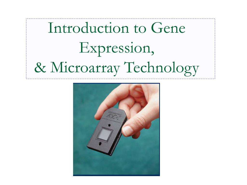
Introduction to Gene Expression, & Microarray Technology. Gene expression. Gene expression is the process by which information from a gene is used in the synthesis of a functional gene product. These products are often proteins. Also known as DNA Chip.
Introduction to Gene Expression, & Microarray Technology - slide 1
01 / 51Introduction to Gene Expression, & Microarray Technology
1 / 17

Télécharger la présentation

Introduction to Gene Expression, & Microarray Technology
Une image / un lien ci-dessous est fourni (tel quel) pour télécharger la présentation

Politique de téléchargement : le contenu du site vous est fourni TEL QUEL pour votre information et votre usage personnel ; il ne peut être vendu, concédé sous licence ni partagé sur d’autres sites sans l’accord de son auteur.Le contenu vous est fourni TEL QUEL, pour votre information et votre usage personnel uniquement. Téléchargez la présentation en cliquant sur ce lien. Lors du téléchargement, si vous ne parvenez pas à télécharger une présentation, l’éditeur a peut-être supprimé le fichier de son serveur.Pendant le téléchargement, si vous ne parvenez pas à obtenir une présentation, le fichier a peut-être été supprimé par l’éditeur.

F I N

Plus de contenu associé

Transcription de la présentation
  1. Introduction to Gene Expression,& Microarray Technology

  2. Gene expression • Gene expression is the process by which information from a gene is used in the synthesis of a functional gene product. • These products are often proteins

  3. Also known as DNA Chip • Allows simultaneous measurement of the level of transcription for every gene in a genome (gene expression)

  4. Are small, solid supports onto which the sequences or subsequences from thousands of different genes are attached. • The supports are usually glass microscope slides, or silicon chips or nylon membranes. The DNA is printed, spotted, or actually synthesized directly onto the support. • The spots can be DNA, cDNA, or oligonucleotides

  5. It consists of an arrayed series of thousands of microscopic spots of DNA oligonucleotides, each containing specific DNA sequence.

  6. DNA microarrays are created by spotting every gene in a genome onto a glass microscope slide.

  7. Each spot represents different gene/clone Modified fromhttp://darwin.bio.uci.edu/~faculty/wagner/array2.html

  8. Why Are Microarrays Important? • Microarrays are a significant advance both because they may contain a very large number of genes and because of their small size. • Microarrays are therefore useful when one wants to survey a large number of genes quickly or when the sample to be studied is small. • .

  9. The process

  10. Sample preparation

  11. The two samples to be compared . • In this example treated sample (case) and untreated sample (control).

  12. DNA probe • A short sequence of DNA labelled that is used for the detection of a complementary nucleotide sequence.

  13. Hybridization cover slip Hybridize for 5-12 hours Binding of cDNA target samples to cDNA probes on the slide

  14. DNA microarrays: step by step • Production of DNA probes • Printing or “spotting” Printing or “spotting”

  15. The arrayer Ngai Lab arrayer , UC Berkeley Print-tip head

  16. Once extracted, the mRNAs need to be labelled with fluorescent markers1 so that they can be detected later, on the surface of the micorarray. • mRNA of the control cells is usually labelled with green fluorescent marker, and mRNA of the cells under study with red fluorescent marker.

  17. the control mRNA (labelled green) is mixed with the test mRNA (labelled red). • The mixture is then flooded over the surface of a slide, which is then incubated at 42°C, • After 12 hours, the microarray is washed . • The microarray is now ready for scanning.

  18. Labeled DNA hybridizes to corresponding DNA/gene

  19. Image Duplicate spots Scanning Detector PMT

  20. But wait a minute there are not just red and green dots there are yellow dots as well!

  21. This can easily be explained. • It is clear that the red spots contain mRNA from cancer cells and green spots mRNA from noncancerous control cells.

  22. yellow! Remember mRNA hybridizes with its complementary DNA and one spot on the microarray represents billions of copies of DNA from ONE gene. • In other words, when a spot is yellow, there are equals amounts of mRNA of the gene found in cancerous and control cells.

  23. And black means that there is no mRNA of that gene either in the control or cancerous cells.

  24. RGB overlay of Cy3 and Cy5 images