Download

1 / 8
80 likes | 103 Vues
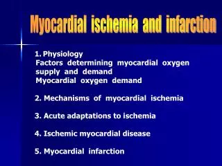
This article explores the causes and mechanisms of inadequate myocardial oxygen supply, including limited flow in epicardial coronary arteries, endothelial dysfunction, impaired microvascular coronary flow reserve, and extravascular compression. It also discusses the association between ischemia and symptoms, non-invasive tests for diagnosis, and the role of coronary microcirculation dysfunction in ischemic heart disease.

E N D
Causes of inadequate myocardial O2 supply Limited flow in epicardial coronary arteries • Flow-limiting lesion(s) in conduit vessels • Exacerbated by impaired endothelium-dependent response to stress • Reduced dilation • Constriction Impaired microvascular coronary flow reserve • Resistance vessel (<200 µm diameter) dysfunction • Disordered VSMC activation/contraction • Abnormal motility • Abnormal growth • Inflammation Extravascular compression Pepine CJ et al. J Am Coll Cardiol. 2006;47:30S-5. Reis SE et al. J Am Coll Cardiol. 1999;33:1469-75. Kerins DM et al. In: Goodman and Gilman’s The Pharmacological Basis of Therapeutics. 10th ed.
Ischemia is related to myocardial O2 supply and demand Heart rate Diastolic time Spasm/ autoreg. Contractility Coronary blood flow Oxygen demand Oxygen supply Collaterals Wall tension AoP – LVED gradient Systolicpressure Volume Ischemia LVEDP Ao dias. pressure Adapted from Morrow DA et al. In: Braunwald’s Heart Disease. 7th ed.
Symptoms occur at end of ischemic cascade Abnormalities evolving during ischemia • Approximately ½ of patients with angina also experience episodes of asymptomatic (silent) ischemia • Many episodes of ischemia never become painful Angina ST Filling Magnitude of ischemia Systolic dysfunction Relaxation (diastolic dysfunction) 0 30 Duration of ischemia (sec) Cohn PF et al. Circulation. 2003;108:1263-77.Adapted from Kern MJ. In: Braunwald’s Heart Disease. 7th ed.
Obstructive plaque and ischemia Obstructiveatheroscleroticplaque Fattystreak Increasedplaque Normal Plaque Exertionalangina Noninvasive tests: normal Noninvasive tests: abnormal Vasodilator response to stress Adapted from Abrams J. N Engl J Med. 2005;352:2524-33.
Impaired microvascular perfusion in the anginal syndrome Diminished microvascular perfusion P < 0.001 P = 0.002 P = 0.02 P = NS Control (n = 10) Chest pain with normal coronary angiogram (n = 20) *Assessed via magnetic resonance imaging Panting JR et al. N Engl J Med. 2002;346:1948-53.
Subendocardial hypoperfusion: Association with anginal syndrome MRI of myocardium during first pass of gadolinium • At rest • During stress (adenosine infusion) Healthy control Patient withchest pain and angiographically normal coronary arteries Magnetic resonance imaging Panting JR et al. N Engl J Med. 2002;346:1948-53.
Accumulating evidence implicates coronary microcirculation dysfunction in IHD • Wide variability in effort tolerance over time • Large scatter between stenosis severity and flow reserve • Reduced flow responses to stress in regions perfused by non-stenotic vessels • Variability in outcome after successful intervention • ~25% of cases with biomarker-positive ACS have no flow-limiting stenosis • Predictive value of BNP and CRP for adverse outcomes in ACS • Plaque erosion with microvascular embolization Pepine CJ et al. J Am Coll Cardiol. 2006;47:30S-5.