Download
1 / 35
350 likes | 570 Vues
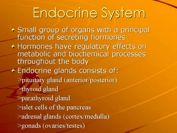
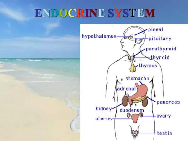
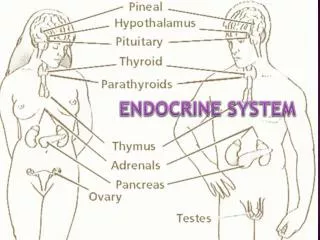
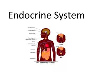
The Physical Basis of Neuronal Function. Major communication systems coordinate parts of animals body Neuronal system: Rapid & Short Burst Endocrine system: Slow & Persistent. Nervous System. CNS (central nervous system): Brain Spinal cord PNS (peripheral nervous system):

E N D
The Physical Basis of Neuronal Function • Major communication systems coordinate parts of animals body • Neuronal system: Rapid & Short Burst • Endocrine system: Slow & Persistent
Nervous System • CNS (central nervous system): • Brain • Spinal cord • PNS (peripheral nervous system): • Cranial nerves (from brain) • Spinal nerves (from spinal cord) • 2 Types of cells in nervous system: • Neurons • Supporting cells
Neurons Neurons (nerve cell): major players in the nervous system; are specialized cells that receive information, process information and transmit the information to other cells;communicate information using combination of electrical and chemical signals. • Structural and functional units of the nervous system. • Respond to physical and chemical stimuli. • Produce and conduct electrochemical impulses. • Release chemical regulators. • Can not divide by mitosis.
Neurons Most neurons are morphologically asymmetric cells 3 Principal regions: • Soma (Cell body): • Contains the nucleus, metabolic maintenance • Dendrites: • Provide receptive area. • Transmits electrical impulses to cell body. • Axon: • Conducts impulses away from cell body.
Functional: Based upon direction impulses conducted. Sensory or afferent: Conduct impulses from receptors to CNS. Motor or efferent: Conduct impulses out of CNS to effector. Association or interneurons: Located entirely within the CNS. Classification of Neurons
Typically, a region of neuron membrane called spike initiation zone integrates signals from many input neurons to determine whether the neuron will initiate its own signals, called action potential. Neurons transmit electrical signals without loss of signal strength (active electrical properties) Neurons possess various type of ion channels
Information is typically carried through a neuronal circuit via electrical action potential alternating with chemical siganls • Afferent neurons • Synapses • Efferent neurons • Interneurons • Neuronal circuit • Presynaptic • Postsynaptic
Membrane potential (Vm): The electronic potential measured from within a cell relative to the potential of the extracellular fluid, which is by convention considered to be zero; the potential difference across a membrane. Resting potential (Vrest): The normal membrane potential of a cell at rest.
Electrochemical Potential • Theoretical voltage produced across the membrane if only 1 ion could diffuse through the membrane. • Magnitude of difference in charge on the 2 sides of the membrane.
Membrane Potential • Proteins and phosphates are negatively charged at normal cellular pH. • These anions attract positively charged cations that can diffuse through the membrane pores. • Membrane more permeable to K+ than Na+. • Concentration gradients for Na+ and K+. • Na+/ K+ATP pump 3 Na+ out for 2 K+ in. • All contribute to unequal charge across the membrane.
Resting Membrane Potential • The normal membrane potential of a cell at rest, lies -30 and -100 mV. • Cell membrane is selectively permeable to some ions • Unequal distribution of inorganic ions between cell interior and cell exterior. • Any ions that cannot cross the membrane have no effect on the membrane potential.
Nernst Equation • Membrane potential that would exactly balance the diffusion gradient and prevent the net movement of a particular ion. • Equilibrium potential for K+ = - 90 mV. • Equilibrium potential for Na+ = + 60 mV.
Voltage Gated Ion Channels • Changes in membrane potential caused by ion flow through channels. • Specific ion channels for Na+ and K+. • Passive channels are always open. • Voltage gated channels open in response to change in membrane potential.
AP followed by increase in Na+ diffusion. After short delay, increase in K+ diffusion. Membrane Permeability
Action Potential (AP; nerve impulse, spike): • Transient all-or-none reversal of a membrane potential produced by regenerative inward current in excitable membranes. • Three key elements for AP • Active transport • Unequal distribution of ions • Electrochemical gradient Overshoot: The reversal of membrane potential during action potential; the period of time during which cell becomes inside-positive. After-hyperpolarization (undershoot): The transient period at end of of action potential when Vm is more negative than Vrest.
Electrical Activity of Axons • Depolarization: • Potential difference reduced (become more positive). • Repolarization: • Return to resting membrane potential (become more negative). • Hyperpolarization: • More negative than resting membrane potential.
Action Potentials (AP) • Stimulus causes depolarization to threshold. • Voltage gated (VG) Na+ channels open. • Electrical and chemical (electrochemical) gradients inward. • + feedback loop. • VG K+ channels open. • Electrochemical gradients outward. • - feedback loop. • Changes in membrane potential constitute AP.
Action Potentials (AP) • Once AP completed, Na+ / K+ ATPase pump extrude Na+, and recover K+. • All or none: • When threshold reached, maximum potential change occurs. • Coding for Intensity: • Increased frequency of AP indicates greater stimulus strength.
Conduction of Nerve Impulses • Cable properties: • Ability of neuron to transmit charge through cytoplasm. • High internal resistance. • An AP does not travel down the entire axon. • Each AP is a stimulus to produce another AP in the next region of membrane with VG channels.
Refractory Periods • Absolute refractory period: • Axon membrane is incapable of producing another AP. • Relative refractory period: • Axon membrane can produce another action potential, but requires stronger stimulus.
Threshold stimulus: The minimum stimulus energy necessary to produce a detectable response or an all-or-none response 50% of the time.
Accommodation Temporary increase in threshold that develops during the course of a stimulus
Phasic response: The response of a neuron that, when stimulated continuously by a current of constant intensity, accommodates rapidly and generates action potentials only during the beginning of the stimulus period Tonic response: The response pattern of neurons that, when stimulated continuously by a current of constant intensity, accommodate slowly and fire repetitively with gradually decreasing frequency.
Molecular structure of voltage-gated K+channel • Oligomeric complex of four monomeric a-subunits • Four b-units associated with each a-subunit • Each a-subunit (70 kD) has six transmembrane domains • S4 is voltage sensor • Molecular structure of voltage-gated Na+channel • One large a-protein (~260 kD)