Ascites
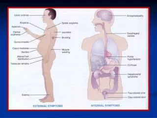
Ascites. Associate Professor Dr Meltem Ergun Yeditepe University Department of Gastroenterology. Points of this lecture. What is ascites? Etiologies of ascites Clinical presentations Pyhsical examination findings Laboratory tests Prognosis Spontaneous bacterial peritonitis.

Ascites
E N D
Presentation Transcript
Ascites AssociateProfessorDr Meltem Ergun Yeditepe University Department of Gastroenterology
Points of thislecture • What is ascites? • Etiologies of ascites • Clinical presentations • Pyhsical examination findings • Laboratory tests • Prognosis • Spontaneous bacterial peritonitis
Definition Pathological accumulation of fluid in abdominal cavity >50 ml
Pathophysiology of Ascites From: Robbins Basic Pathology
Imaging • Ultrasound with Dopplers • Easily confirms ascites • May see nodularity of cirrhosis • Evaluate patency of vasculature • No radiation, contrast • CT / MRI • Evaluation for malignancy
Serum to Ascites Albumin Gradient • Is portal hypertension present? • 97% accurate SAAG > 1.1 g/dL Portal HTN SAAG < 1.1 g/dL Other causes The serum-ascites albumin gradient is superior to the exudate-transudate concept in the differential diagnosis of ascites. Runyon BA; Montano AA; Akriviadis EA; Antillon MR; Irving MA; McHutchison Ann Intern Med 1992 Aug 1;117(3):215-20.
Cell Count, differential and culture • Is ascites infected? • Greater than 250 PMN = SBP • If ascites is bloody ( > 50,000 RBC/mm3), correct by subtracting 1 PMN / 250 RBC • Is ascites bloody? • 5% of pts w/ cirrhosis - spontaneous or s/p traumatic tap. • Non-traumatic associated with malignancy • 20% of malignant ascites • 10% of peritoneal carcinomatosis
Total Protein • Exudate ( > 2.5 g/dL) or Transudate? • Supplanted by SAAG • Is there gut perforation? (vs SBP) • Total protein >1 g/dL • Glucose <50 mg/dL (2.8 mmol/L) • LDH greater than serum ULN
Glucose and LDH • Consistent with infection or malignancy? • Infection and cancer consume glucoselow • LDH is a larger molecule than glucose, enters ascitic fluid with difficulty. • Ascitis/Serum LDH ratio • ~ 0.4 in cirrhotic ascites • Approaches 1.0 in SBP • >1.0, usually infection or tumor
Other tests • Amylase • Uncomplicated cirrhotic ascites • About 40 IU/L. The AF/S ratio is about 0.4 • Pancreatic ascites • About 2000 IU/L. The AF/S ratio is about 6 • Triglycerides — run on milky fluid. • Chylous ascites - TG > 200 mg/dL, usually 1000 mg/dL • Bilirubin — run on brown ascites. • Biliary perforation – AF Bili > serum Bili
Tests for TB • Smear – extremely insensitive • Culture – 62-83% when large volumes cultured • Cell count – mononuclear cell predominance • Adenosine deaminase – • Enzyme involved in lymphoid maturation • Falsely low in pts with both cirrhosis and TB
Cytology • “almost 100%” with peritoneal carcinomatosis have positive cytology • Malignant ascites from massive hepatic mets, HCC, lymphoma are usually negative • Overall sensitivity for detection of malignancy-related ascites is 58 to 75 %
Not helpful • “Some tests of ascitic fluid appear to be useless. These include pH, lactate, and ‘humoral tests of malignancy’ such as fibronectin, cholesterol, and many others”
Biopsy Fatty Liver Cirrhosis http://library.med.utah.edu/WebPath/LIVEHTML/LIVERIDX.html#2
Malignant Ascites • Definition: abnormal accumulation of fluid in the peritoneal cavity as a consequence of cancer. • Commonly caused by cancers of: • Breast, bronchus, ovary, stomach, pancreas, colon • 20% of cases have tumors of unknown primary • Survival poor – usually less than 3 months Becker, G. Malignant ascites: Systematic review and guideline for treatment. European Journal of Cancer 42 (2006) 589 - 597
Malignant Ascites: Pathophysiology • Obstruction of lymphatics by tumor • Prevents absorption of fluid and protein • Alteration in vascular permeability • Hormonal mechanisms (VEGF, IL2, TNF alpha) • Decreased circulating blood volume • Activates RAAS leading to Na retention
Management of Malignant Ascites • Therapeutic paracentesis • Removing up to 5L appears safe • No good data on role of volume expanders • Diuretics • Equivocal evidence of efficacy • May be helpful for portal HTN • Less/minimally useful when no portal HTN • Drainage Catheters • Peritoneovenous shunts
Peritoneovenous Shunt Contraindications • Protein > 4.5 g/l (occlusion) • Loculated ascites • Coagulopathy • Advanced renal/cardiac disease • GI malignancy Complications • Infection • Hematogenous spread of mets • DIC • Pulmonary edema • Pulmonary emboli Denver Shunt (Similar to LaVeen Shunt)
66 yrs old, man presented with abdominal distension and jaundice , started 3 months ago. He had been diagnosed HBS Ag carrier 20 years ago but had no follow up.
Examination of Abdomen • Percussion • liver • Spleen • Palpation • Liver • Spleen • Examination of ascites
Radiological findings • Abdominal ultasonography • Irregular and nodular surface of the liver • Splenomegaly • Ascites • Abdominal MRI and CT • Sometimes better than USG
How about liver biopsy • Itshighlyinvasive • %1-2 severe bleedingand sometimesmortality • Hematologicproblems • Sowhenbiopsy? • ifclinically, radiologicallyandlaboratuaryfindings do not clearlyindicatecirrhosis • Ifwe do not clarifytheetiologicfactorandsuspect of treatablecondition
Staging of Cirrhosis Interpretation:Class A: 5-6Class B: 7-9Class C: 10-15
Prognosis of Cirrhosis Mortality is usually related with the occurence of complications
Treatment At Early Stage • Early treatment may affect prognosis • Stop alcohol drinking • Eradication of viruses • Treatment of the cause • Autoimmune • Wilson • Hemachromatosis • Sclerozing Cholangitis • Very good followup • Prevention of complications • Low Na Diet
Treatment for Ascites • Diuretics (Spironolactone, furosemide) • Low sodium diet • Therapeutic parasentesis • Transplantation
Ascites; usuallythefirstcomplication !! • Sometimes no symptom • Abdominal distension • Feeling abdominal tenderness • Treatment • Na restriction • Dıuretics • Terapeutic parasentesis
Serum alb – ascites alb >1.1 • LDH, protein, glucose • Cell count • Cytology • Culture
Spontaneous bacterial peritonitis • Spontaneousinfection of ascites • Anycirrhoticpatientwith • Fever • Abdominalpain • Abdominaltenderness • Detoriation of clinicalsituation • Parasentesis • Neutrophilcount>250/ml • Ascitesculture: mostly E.coli • Treatment • i.v antibiotics: 3th generationcephalosporins (cefotaxim) firstchoise, • quinalonsorpenicilins
Spontaneus Bacterial Peritonitis: Mechanism Abdominal Cavity Ascitic Fluid Systemic Circulation Intesine • Loss of opsanization • Decreased complemant amount Decreased RES Function Intestinal Permeability Bacterial Overgrowth LIVER Collaterally pass Kupfher Cell Loss Portal Vein
Liver Transplantation • Each patient who has the complications must be listed for transplantation • Child Score > 9 • MELD Score > 10 • Urgent Tx • Acute fulminant liver failure • Acute on chronic liver failure
Cadaveric Tx • Living donor Tx • 600-700 Tx each year in Turkey • Mostly living donor • Survival % 80 in 2 years, % 70 in 5 yrs • Immunsupressive treatment after Tx