Innate Immunity
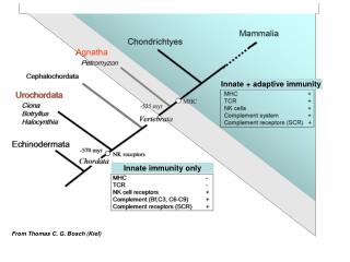
Innate Immunity. Innate Immunity. Mechanisms that do not depend on prior exposure to the pathogen Have evolved over time to protect against groups of organisms that have distinctive molecular features Some elements have also been co-opted for use in the specific acquired immune response.

Innate Immunity
E N D
Presentation Transcript
Innate Immunity • Mechanisms that do not depend on prior exposure to the pathogen • Have evolved over time to protect against groups of organisms that have distinctive molecular features • Some elements have also been co-opted for use in the specific acquired immune response
Elements of Innate Immunity • External Barriers • Phagocytic cells • Complement • Humoral mechanisms • Extracellular Killing
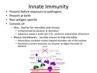
External Barriers • Skin • Very effective barrier to infection – cuts, burns etc, infection is major complication • Mucus and mucosal surfaces • Mucus can trap and move foreign things along • Some secretions have bacteriocidal components • Normal bacterial flora can play a protective fole • Environment (e.g: vaginal pH) • Colicins
Phagocytic Cells • Polymorphonuclear Leukocytes (a.k.a PMNs or “Polys” • Macrophages (lit: “big eater” Gr.)
Phagocytic Cells • Polymorphonuclear neutrophils: PMNs • White cells (leukocytes), multi-lobed nuclei (look like many nuclei in section) • Neutrophil: does not take up the major dyes • Short-lived; lots of glycogen (energy from glycolysis even if O2 is not available) • Macrophages • Mononuclear cells • Long lived, significant RER • Do not circulate, but hang out in lymph nodes and other interesting places
Recognition by Phagocytic cells • Phagocytes have evolved a system of receptors that can recognize molecular patterns on the surface of pathogen that are • Conserved • Shared by many infections agents • Different from “self”-patterns • Examples: • The lipopolysaccharide of certain bactreia • Yeast cell wall mannans • Mycobacterial glycolipids • But not glactose and sialic acid group that are typical the ends of mammalian surface polysaccharides
Phagocytosis • Engagement of receptors generates internal signal to start phagocytosis • An actin—myosin contractile sytsem for putting pseudopods around the particle • Lysosomes discharge their contents into the phagosome • Several biochemical pathways ensue to degrade content
Complement • A system of 20 or so different proteins that are “set off” in a cascade of events. • Cascade: one molecule, when activated, catalyzes a change in the next, makes it active, so that it catalyzes a change in the next etc. Such protein cascades allow a profound response to a trigger. • Complement proteins modify each other, and the fragments may have other effects as well
Complement has 2 pathways: the classical and the “alternative” • The classical pathway was figured out first, but may be more recent in evolution, because it depends acquired immune response (antibodies) • So we will talk about that later! • The “alternative” pathway is activated by microbial polysaccharides, and is part of innate immunity
Complement: Act I, Scene 1Complement activation by bacterial cell walls • C3 is routinely cleaved at slow rates to C3b • Factor B is cleaved by a plasma enzyme, Factor D to Bb • C3b and Bb bind together to form an enzyme, C3bBb, or C3 convertase. • This convertase very actively cleaves C3 to C3a and C3b • C3b is the central molecule – sets off complement-mediated killing ….. so how is this prevented from running amok in the uninfected state? • A protein H binds to C3bBb, and then another protein I binds to the C3bBbH complex, inhibiting it. However ….
Complement: Act I, Scene 2If certain bacteria are present, the cascade is not inhibited. • Some mico-organisms can stabilize the convertase C3bBb, so that it does not bind Factor H • If the convertase does not bind H, it also does not bind I, so it is not inhibited, so…. • The reaction does get amplified, and much more • C3b is made
Complement: Act II • The C3b can for a short time, react covalently with local hydroxyl or amino groups at the microbial cell surface • C3b bound to the surface makes the cell “tasty to macrophages” – the surface is “opsonized” • In the presence of extra C3b, the C3bBb changes its enzymatic specificity such that it now cleave C5 into C5a and C5b • C5b stays bound to C3b at the surface of the invading cell. • Seeing C5b bound, C6, C7, C8 and C9 bind, and form a membrane pore (called the Membrane Attack Complex) • The cell lyses
Complement: Act III • The smaller fragments C3a and C5a stimulate • Phagocytosis • Mast cell degranulation • Histamine (causes vasodialation, capillary permeability, bronchioconstriction) • Proteases and other degradative enzymes • Chemotactic factors (recruit phagocytes) • Interleukins further activation of macrophage activity , and other things = basic inflammatory response
Humoral Mechanisms provide a second defensive strategy • The term “humoral” means soluble in serum of other fluids; non involving cells • Antibodies are humoral defenses (but specific acquired immunity, so discussed later) • Among the innate humoral immune defenses: • Complement • Clotting Factors (cascade, fibrin and fibinogen) • Lysozyme (in many secretions, attacks bacterial cell wall) • Acute phase proteins • Intereferons
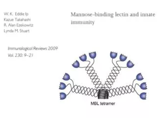
Acute Phase Proteins • Concentration increases in response to alarm proteins, such as IL-1 • IL-1 is released by macrophages when they are activated (by bacterial endotoxins, foreign molecular patterns, complement etc) • Increase in C-Reactive protein, and mannan binding proteins (as much as 1000-fold). • These bind to bacteria, and activate the classical pathway of complement (take the place of the C1 proteins that are normally activated by Ab) • C3b opsonizes the foreign cells
Extracellular Killing • NK (Natural Killer) cells kill virally- infected cells using perforin • Induce apoptosis • Apoptosis = Programmed cell death • A series of reactions, cascade of proteolytic enzymes (capases); the end result is rapid degradation of the nucleus, and cutting DNA in to nucleosome size pieces