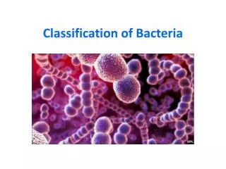
Classification of bacteria
Classification of bacteria. Kingdom: Monera Division: Eubacteriophyta Class: Bacteria. What is bacteria?. They are prokaryotic organisms : (they Don’t have organized nucleus surrounded by nuclear membrane and the DNA found free in the cytoplasm).

Classification of bacteria
E N D
Presentation Transcript
Classification of bacteria • Kingdom: Monera • Division: Eubacteriophyta • Class: Bacteria
What is bacteria? • They are prokaryotic organisms: (they Don’t have organized nucleus surrounded by nuclear membrane and the DNA found free in the cytoplasm). • organisms made up of just one cell. • capable of multiplying by themselves, as they have the power to divide , this is called ( binary fission). • some bacteria can cause diseases.
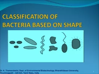
Common Shapes of Bacteria BACILLUS COCCUS vibrio SPIRILLUM
Aggregation of coccus shape: monococcus Staphylococci
Aggregation of Bacillus shape monobacillus
Culture medium • Is a mixture of various nutrients which are suitable for the growth of microorganisms • At least 500 different types • Solid or liquid • Inoculated by loops, needles, pipettes, and swabs
Types of media Based on the physical state • Liquid medium: • Without agar. • for the proliferation of bacteria. • Solid medium: • 1.5-2.5% agar. • for the isolation and identification of bacteria • e.g., slant, Petri dishes. • Semisolid medium: • 0.3-0.5% agar. • for the observation of bacterial motility and preservation of bacteria.
Semisolid media Liquid media
Where we can find and isolate Bacteria? • We can isolate bacteria from any source: • Air • Water • Dust • Human body ex.“skin, mouth and nails” • Foods • Any other sources
Colony morphology • Shape of colony • Edge • Elevation
Staining of Bacteria • Bacterial cells are almost colorless and transparent • A staining technique is often applied to the cells to color them→ Their shape and size can be easily determined under the microscope. • Staining may be a simple stain ( use only one type of stain ex: methylene blue) or complex stain ( ex: Gram stain)
Smear Preparation: Preparation and Fixation of Bacteria for Staining. Objective: To kill the microorganism & fix them to the slide to prevent them from being washed out during the process of staining.
Smear preparation • Flame the loop and let it to cool. • Put the bacterial suspension on a clean slide. • Fix the bacterial suspension by flam (avoid overheating).
Gram Stain: It is the most important differential stain used in bacteriology because: it classified bacteria into two major groups: • Gram positive: • Appears violet after Gram’s stain b)Gram negative: Appears red after Gram’s stain
Procedure: Crystal violet (30-60 sec) ↓wash with water Iodine (2 min) ↓ wash with water Alcohol (10 sec) ↓ wash with water Safranin (1 min) wash with water, dry and examine with oil lens
Results: Shape: Cocci Aggregation: irregularclusters Colour: Violet Gram’s reaction: Gram’s+ve Gram positive Staphylococci
Results: Shape: Bacilli Aggregation: Single Colour: red Gram’s reaction: Gram’s –ve Gram negative monobacilli