Download
1 / 16
170 likes | 400 Vues
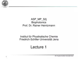
ASP_MP_S2j Biophotonics Prof. Dr. Rainer Heintzmann. Institut für Physikalische Chemie Friedrich-Schiller-Universität Jena. Lecture 1. Content. Introduction Contrast modes in light microscopy 2.1 Bright field microscopy 2.2 Dark field microscopy 2.3 Phase contrast microscopy

E N D
ASP_MP_S2j Biophotonics Prof. Dr. Rainer Heintzmann Institut für Physikalische ChemieFriedrich-Schiller-Universität Jena Lecture 1
Content • Introduction • Contrast modes in light microscopy • 2.1 Bright field microscopy • 2.2 Dark field microscopy • 2.3 Phase contrast microscopy • 2.4 Polarisation microscopy • 2.5 Differential interference contrast • Optical coherent tomography • Molecular many electron systems:electronic and nuclear movement • UV-Vis absorption • 5.1 Franck-Condon principle • 5.2 Electronic chromophores • 5.3 Polarimetry & circular dichroism • Fluorescence spectroscopy • 6.1 Stokes shift • 6.2 Fluorescence life time • 6.3 Fluorescence quantum yield • 6.4 Steady state fluorescence emission • 6.5 Fluorescence excitation spectroscopy
Content • Fluorescence microscopy • 7.1 Fluorochromes • 7.2 Confocal fluorescence microscopy • 7.3 FRET • 7.4 FRAP, iFRAP, FLIP • 7.5 Ultramicroscopy / SPIM / HILO • 7.6 Multi-photon microscopy • 7.7 4Pi microscopy • 7.8 STED microscopy • 7.9 linear and nonlinear structured illumination • 7.9 PALM/STORM • Vibrational microspectroscopy • 8.1 Normal modes • 8.2 IT-absorption microspectroscopy • 8.3 Raman microspectroscopy • 8.4 Protein structure determination • 8.5 Biomedical diagnostics • 8.6 Resonance Raman spectroscopy • 8.7 SERS
Content • Non-linear Raman microspectroscopy • 9.1 Hyper Raman • 9.2 Coherent anti-Stokes Raman scattering (CARS) • 9.3 Stimulated Raman microscopy • Future trends in non-linear microscopy
Sciences Biology Physics Chemistry Medicine (wealth of disciplines) Bio-photonics Engineering Optical Engineering Medical Engineering 1. Introduction Biophotonics a highly interdisciplinar approach
1. Introduction Light-Matter Interactions as the basis for Biophotonics
Absorption Scattering Reflection Refraction 1. Introduction: Light-Matter Interactions Light-Matter Interactions incident light reflected light tissue transmitted light scattered light a(n) =absorption cross-section aS = scattering cross-section I(z) = intensity in depth z I0 = incident intensity I(n) = transmitted intensity
1. Introduction: Light-Matter Interactions blood melanosom aorta water skin epidermis
- + E 1. Introduction: Light-Matter Interactions + Polarisation P : Dipole moment per unit volume
1. Introduction: Light-Matter Interactions Linear Polarisation
1. Introduction: Light-Matter Interactions Nonlinear Polarisation for convergence:
1. Introduction: Light-Matter Interactions Nonlinear Polarisation yields:
1. Introduction: Light-Matter Interactions Example Terms in P: Frequency Name DC polarizability optical polarizability (refractive index) DC hyperpolarizability linear electrooptic effect(Pockels Effect) DC hyperpolarizability second harmonic generation third harmonic generation Kerr effect (n=n0+n2I)
c(1) c(2) c(3) • Linear absorption • Spontaneous emission (Fluorescence) • Reflection • Elastic scattering • Inelastic scattering: Raman-scattering • Diffraction • Second harmonic generation (SHG) • Sum-frequency generation (SFG) • Difference-frequency generation (DFG) • Optical parametric amplification • Third harmonic generation (THG) • Two-photon absorption (TPA) • CARS (Coherent Anti-Stokes-Raman-Scattering) 1. Introduction: Light-Matter Interactions Process
2. Contrast modes in light microscopy : 1D monochr. wave Absorption Dispersion nR : real part of refractive index nI : imaginary part of refractive index Phase difference Amplitude difference Refractive indices Wavelength l • Dark field • Phase contrast • Differential phase contrast • Bright field
2. Contrast modes in light microscopy: Bright field • 2.1 Bright field transmission (absorption = imaginary part of refractive index) • An object, keeping the phase of an incoming wave constant and decreasing the amplitude is called amplitude object. • Contrast is A0 –A1,2 • Bright filed microscopy is the most simpleand basic light microscopy method • Sample is illuminated from belowby a light cone • In case there is no sample in the opticalpath a uniform bright image is generated • An amplitude object absorbs light at certain wavelengths and therefore reduces the amplitude of the light passing through the object Amplitude difference Wavelength l Uniform bright field image Bright field image of Moss reeds