Exploring Cell Biology: Microscopy Techniques and Protein Studies
Dive into the fundamentals of cell biology with a focus on microscopy techniques, cellular components, animal tissues, and protein function studies. Learn about different methods to study individual cells and proteins. Discover the key questions and concepts in this fascinating field.

Exploring Cell Biology: Microscopy Techniques and Protein Studies
E N D
Presentation Transcript
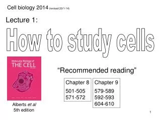
Cell biology 2014 (revised 20/1-14) Lecture 1: How to study cells “Recommended reading” Chapter 8 501-505 571-572 Chapter 9 579-589 592-593 604-610 Albertset al 5th edition
The tree of life Microbiology Microbiology & Cell biology (prokaryotes) Nucleus Eubacteria Eukaryotes Archaea Cytosol
Biology Cell biology Molecular biology Organism biology Met Ser Arg Pro Nanometers Micrometers Millimetres Meters
I am seeing atoms The starting point of cell biology: microscopy Let's call them cells (1665) Robert Hooke (1635 – 1703) Cellulae, little room Sliced cork
Conceptual breakthroughs in cell biology Mikroskopische Untersuchungen über die Übereinstimmung in der Struktur und dem Wachsthum der Tiere und der Pflanzen (1839) - All organisms consist of one or more cells - The cell is the basic unit of structure Die Cellularpathologie (1858) - All cells arise from preexisting cells On the Origin of Species by Means of Natural Selection (1859) - All cells have a common ancestor Zellsubstanz, Kern und Zelltheilung (1882) - Chromosome (thread) segregation during mitosis (i.e. precise partitioning/transport of defined cell structures)
All eukaryotic cells are in principle very similar - Organelles - Cytoskeleton - Nucleus - Chromosomes • Key questions in cell biology • Structure and functions of cellular components • How do cells communicate? • Which signals trigger cell cycle entry? • How is cell duplication coordinated? • How is one cell split into two?
Multicellular eukaryotes – not just cells The extra cellular matrix (ECM) works as a scaffold in metazoans supporting cells in various ways
Animal tissues mainly consisting of (different) cells - Epithelia Protective covering of surfaces, both outside and inside the body - Muscle Force generating cells (contraction) Animal tissues consisting of cells and ECM - Connective • Hard tissues of bone and teeth • Transparent matrix of the cornea • Ropelike organization of tendons
How to study individual animal cells Primary cell cultures Secondary culture Explants Proliferation (growth factors) Complete tissue section Only cells Immortalization (e.g. by oncogenes) Cell line, with indefinite proliferative potential Tumor patient
I. How to study the function of a protein in cells Depletion/mutation of endogenous protein Normal (Control) Overexpression of protein (ectopic expression)
II. How to study the function of a protein in cells Central dogma of molecular biology • Loss-of-function mutations • Gain-of-function mutations • Overexpressed (trans)gene DNA Transcription - RNA interference mRNA Translation • Inhibitory (pharmaceutical) drugs • new field ”chemical genetics” Protein
RNA interference – depletion of a specific protein ds short RNA (synthetic or expressed as shRNA) RISC mRNA Duplex formation mRNA detroyed Normal cell RNAi treated cell DNA: mRNA degraded! mRNA: Protein: Already existing proteins decay over time
Systems for overexpression of a protein Transient transfection (plasmid DNA is not replicated) Stable transfection (Chromosomal integration) drug resistance Plasmid + Quick (4 – 6 hours) High expression level + Homogenous cell line Unlimited amount of transfectants • 4 – 6 week to establish a cell line • Impossible if gene product • causes a cell cycle block • Heterogeneous cells • Small amount of transfectants
The development of microscopy ~1900 Today Zacharias Janssen (1580 -1638) The first microscope
The three principle tasks of microscopy - Produce a magnified image (magnification) - Separate the details in the image (resolution) Resolution: the smallest distance between two objects at which the two objects can be seen as separate units Maximal resolution = l/2 - Render the details visible (contrast)
Bright field microscopy Ocular Objective Stage Condenser Lamp
Specialized bright field microscopy Enhances the contrast between intracellular structures Bright field Phase contrast Differential interference contrast (DIC)
Creation of contrast in bright field microscopy Unstained cell Stained cell Classical stains
Preservation of biological structures by fixation Process in which cellular structures are preserved and fixed in position by chemical agents Formaldehyde Glutaraldehyde Alcohols Extensive protein cross-linking Protein denaturation Fixation may introduce structural artifacts
Shortcoming of bright field microscopy Okay this was interesting..... ...but where is the protein of interest?
1. 2. Raising antibodies against specific proteins Polyclonal antibodies Purify antibodies from the blood of the animal Epitope Protein X Monoclonal antibody Take out antibody producing B cells Protein X + Molecmodels. 25.2-antibodies Fuse with myeloma cell to generate a hybridoma
Detection of specific proteins with antibodies Secondary antibody Specific to the primary antibody, conjugated with e.g. a fluorochrome Primary antibody Specific to epitope on protein X Protein X Protein X Protein X The primary antibody (e.g. rabbit) is recognized by many secondary antibodies (e.g. goat anti-rabbit) Signal amplification
Principle behind a fluorochrome Fluorochrome Excitation Emission - - - - - - - - Fluorochrome # 1 Fluorochrome # 2 A fluorochrome absorb light of a particular wavelength and re-emit light of a longer wavelength
How it works in reality Emission filter Filter cube Excitation filter Beam splitter - - - -
e- e- Electron microscopy (EM) Maximal resolution = l/2 400 700 nm Maximal resolution 200 nm Resolving smaller structures demands something with a much shorter wavelength + 100 000 V l= 0.004 nm Resolution 0.002 nm (0.1 nm in reality)
e- e- Transmission Electron Microscopy (TEM) Electron gun Vacuum! Supporting grid Very thin section of a cell stained with heavy metal Detector
e- e- e- e- Detector Scanning Electron Microscopy (SEM) Visualizing surface features The specimen is coated with metals to deflect electrons Sequential scanning Electron gun Cell with metal coating
Different forms of microscopy Bright field microscopy Fluorescence microscopy cell organelles Location of molecules large molecules Different techniques – different ”windows” Electron microscopy
The fluorescent protein revolution YFP DsRed GFP Aeqourea victoria Protein X GFP Protein X GFP - - Transient or stable expression Detection in either live or fixed cells Video 02.3-brownian_motion.mov Video 10.6-FRAP - - Visualization of signaling in live cells (NFAT): Video 12.2-nuclear_import.mov