Download
1 / 32
320 likes | 426 Vues
Understand lesion localization in neurology with this informative guide. Learn about ALS, CST, reflexes, and more for clinical success.

E N D
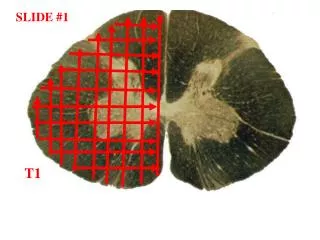
SLIDE #1 T1
SLIDE #3 PONS
A SLIDE #12 C B D E F
SLIDE #17 D C E A B
SLIDE #18 right ear –light reflex (Lr) is always anterior ventral quadrant Hm=head of malleus (don’t sweat the other abbrevs/labels!)
SLIDE #23 B A E C D
SLIDE #24 G A B C D E F
SLIDE #25 A E D B C
SLIDE #26 A B C D
SLIDE #28 B A
SLIDE #30 C B A
SLIDE #31 • SPEED PLAYS • if both strength and pain/temperature are impaired in a single limb, the lesion is either in the periphery or the cortex. The ALS carries crossed info in the spinal cord while the LCST controls ipsi muscles via LMNs. The ALS and CST are physically separated as they go up the brain stem, so usually are not damaged together. However, they are pretty close in cortex (pre- versus postcentral gyrus) and could be damaged at the same time. • if there is reduced pain/temperature in one limb and reduced vibration sense in a contralateral limb, the level of the lesion is somewhere in the spinal cord (on the side of the vibration/position loss) • increased reflexes in a symptomatic limb (after spinal shock) suggests a central (CNS/spinal cord) lesion; reduced reflexes in a symptomatic limb suggest a peripheral lesion • facial weakness ipsilateral to body weakness implies a lesion rostral to motor VII (rostral pon and above) • both a third nerve palsy and a Horner’s can result in ptosis and pupillary asymmetry—but with a third nerve palsy the ptosis is on the side of the large pupil; with a Horner’s the ptosis is on the side of the small pupil • 6. diplopia is always due to a lesion in the brain stem or periphery (nerve/NMJ/muscle) but not in the cortex (frontal eye fileds). A gaze palsy (impaired movement of both eyes in one direction, but both eyes move congruently and remain aligned in all postions of gaze) is due to a lesion in cortex or brain stem, but not in the periphery
SLIDE #32 I guess this is it!! Sorry to see it end!!