Download
1 / 25
250 likes | 291 Vues
This guide covers preoperative considerations, postoperative management, nursing diagnoses, interventions, complications, and catheterization in kidney surgery. It includes detailed information on patient assessment, monitoring potential complications, and catheterization purposes and complications.

E N D
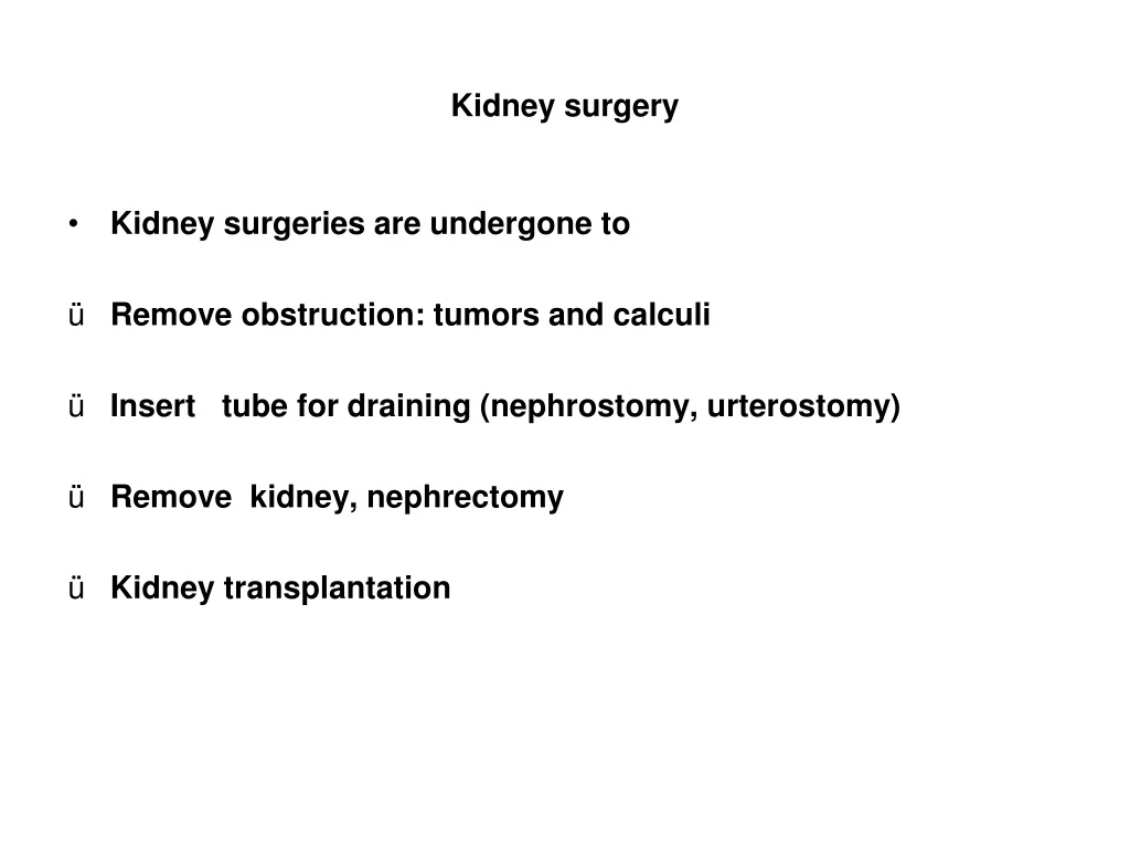
Kidney surgery • Kidney surgeries are undergone to • Remove obstruction: tumors and calculi • Insert tube for draining (nephrostomy, urterostomy) • Remove kidney, nephrectomy • Kidney transplantation
Preoperative consideration • Assess and maintain renal function • Encourage fluid intake to remove waste products, unless contraindicated • Manage infection, antibiotics with extreme care because they are toxic • Coagulation studies: PT, PTT, Platelet counts, if has history of bleeding • Assist patients to express concerns; reinforce confidence • Perioperative concerns: • Flank approach; Lumber approach; Thoracoabdominal approach (figure 44-10, P. 1553) • Read Nursing Care Plan for Kidney Surgery. Chart 44-9, P. 1555-1558.
Postoperative management • Immediate postoperative care • Respiratory status; Circulatory status; Pain • Hemorrhage and shock are chief complications • Fluid & blood replacement is necessary immediately after surgery; to manage intra-operative loss • Abdominal distention & paralytic ileus are common due to: reflex paralysis of peristalsis; manipulation of colon • Relieved by decompression through nasogastric tube • Oral fluid are permitted when passage of flatus is noted • Infection, culture and antibiotic; consider nephrotoxicity • Low-dose heparin, to prevent thromboembolism
Postoperative assessment • Assess respiratory • Location of incision-causes pain on inspiration & coughing • Lung sounds, rate, pattern of breathing • Cardiovascular status: • VSs arterial blood pressure CVP • Skin color & temperature, urine out put • Surgical incision & drainage tubes for evidence of bleeding • Pain: incisional pain, abdominal distention--discomfort • Patency and adequacy of urinary drainage system • Decreased or absent drainage should be promptly reported
Nursing diagnoses • Ineffective airway clearance • Ineffective breathing pattern • Acute pain • Urine retention related to pain, immobility, anesthesia • Potential complications • Bleeding • Pneumonia • Infection • Fluid disturbances • Deep vein thrombosis
Nursing interventions • Maintaining airway clearance and breathing pattern • Relieve pain: Patient controlled analgesia • Promoting urinary elimination • Monitor urine output & drainage • Accurate record fluid from each tube separately • Strict asepsis when manipulating tubes, drainage system • Hand hygiene before touching any part of the system • Use closed drainage system to avoid contamination • Assess drainage volume, color; urine analysis and culture • Urinary bag below bladder and off the floor; irrigation performed carefully with a sterile solution
Monitoring & managing potential complications • Bleeding: monitoring; suspected if urine output is less than 30 ml/hr and fatigue • Pneumonia—incentive spirometer • Infection: prevention and monitoring • Fluid imbalance: observe for Wt gain, edema, urine below 30 ml /hr, adventitious sound • Fluid excess—fluid restriction; give lasix; dialysis may be needed • DVT: elastic compression stockings, monitoring symptoms of thrombosis; leg exercise, heparin • Read Chart 44-12, PP. 1348-1350
Catheterization Purpose • Relieve urinary tract obstruction • Assist with postoperative drainage • Mean to monitor accurate urine output • Promote drainage in patients with NB dysfunction or urine retention • Prevent urinary leakage in stages III, IV of pressure ulcer Complications: • UTI • Bladder spasm • Uretheral stricture • Pressure necrosis
Catheterization • Indwelling catheter; • Use closed drainage system • In transuretheral prostate surgery; the most common is triple lumen catheter; attached to sterile closed drainage system • Bacteria may enter from the port of the urinary bag; keep the bag below the bladder, minimize the risk of contamination • Suprapubic catheter: A measure to divert urine from urethera • To remove: clamp the catheter for 4 hrs; patients void; measure residual; if less than 100ml in 2 occasions, morning and evening; remove catheter • Require fluids to prevent encrustation, wound-ostomy care • Problems: bladder stone, infection,
Nursing management during catheterization Assessing patient and the system • Assess drainage system, color, odor, and volume of urine • Accurate intake & output • Observe the catheter position, no necrosis • Assess for signs of UTI infection; hematuria, fever, anorexia in high risk population Prevent infection • Gentle cleansing to remove encrustation • Fluid intake • Urine culture; • see chart 45-10 P. 1374
Nursing management Minimizing trauma • Use appropriate size • Adequate lubrication • Insert far in the bladder • Secure the catheter properly • Minimize manipulation • Prevent pressure on urethera • Consider confused patients
Nursing managementbladder retraining • During catheterization: • Detrusor does not actively contract the bladder • Detrusor does not respond to bladder filling, when catheter is removed—result in retention or incontinence • Known as post catheterization detrusor instability • management: Retraining of the bladder; void every 2-3 hrs; • Then measure residual urine; using portable ultrasonic bladder scanner; if not completely empty—immediate cath; take few days; see chart 45-10, P1589. • Intermittent self-catheterization in spinal cord injury • Patient teaching: Use of septic technique • Every 4-6 hrs, and just before the bed time. Read p1375.
Lower urinary tract infection • Bladder sterility is maintained by • Physical barrier of urethera; urine flow • Competence of Ureterovesical junction • Antibacterial enzymes & antibodies • Antiadherent effects of mucosal cells of bladder • Infection • Bacteria must gain access and colonize epithelium of the urinary tract, to avoid washed out with voiding • Many UTIs results from fecal organisms that ascend from perineum
UTI • Glycosaminoglycan, a hydrolic protein, has nonadherent protective effect, it forms a water barrier that serves as a defensive layer between urine and the bladder • Urethrovesical reflux: backflow of urine to the bladder—by negative pressure; effect of sneezing • Ureterovesical reflux, when impaired, bacteria may reach kidneys • Uropathogenic: Bacteriuria is 105 colonies of bacteria per milliter of urine—clean-catch midstream—distinguishes bacteriuria from contamination in women • In men, bacteriuria is 104
LUTI • ROUTES OF INFECTION • Transuretheral, ascending infection, most common—fecal contamination, sexual intercourse • Hematogenous, blood stream • Dierect extension, fistula from intestine • Assessment / Clinical manifestations: • 50% report no symptoms; in noncomplicated • Cystitis: Burning urination; frequency; urgency; nocturia • Incontinence; suprapubic or pelvic pain, hematuria; • Complicated: may develop septic shock • Urine culture
LUTI management • Acute pharmacological therapy • Antibiotics with minimal effects on fecal or vaginal flora • Thereby; minimizes the incidence of vaginal yeast infection; yeast viginities • The trends is to shorten the course of therapy, an average 3 days. • In institutional case 7-10 days, in complicated Cephlosporin • Read p. 1574 for antibiotcs use in UTI • Instruct to take the doses prescribed • Long-term doses for pyelonephritis, • hospitalizationand IV are occasionally necessary
LUTI management • Long term pharmacological therapy: • With short-term therapy relapse may occur in 20%; • relapse may be due to • upper UTI , source of infection • Treatment is inadequate; • Administered for a short period • Reinfections with new bacteria occurs in 90% in women
Relapse- management • If no pathological abnormalities: • Women is instructed to use antibiotics on their own when symptoms occurs and; To consult healthcare providers • When symptoms persist; Fever occurs • No. of treatment episodes exceeds 4 in 6 months • If infection recurs after completing the course, another short course (3-4 days) of full dose, followed by a regular bedtime dose • If no recurrence, every other night for 6-7 months • Preventive therapy after sterilization of urine, trimethoprim at bedtime • Inconclusive evidence about the effect of cranberry juice
LUTI Nursing Interventions • Relieving pain • Relieved once antibiotics are initiated; • Antispasmodic to relieve bladder irritability • Analgesics and application of heat to the perineum • Liberal amounts of fluids, water, to promote blood flow and wash bacteria • Avoid urinary tract irritants, alcohol, coffee, tea, cola • Encourage frequent voiding, every 2-3hrs
LUTI Nursing Interventions • Monitoring and managing potential complications • Goal of early treatment is to prevent renal failure; and other complications: urosepsis, strictures, obstructions • Teach a prompt recognition of early symptoms; test for bacteriuria, initiate medications as prescribed • Avoid urinary catheter; if necessary specific nursing intervention to prevent infection p. 1577 • Assess vital signs, blood test culture, WBCs
Upper urinary tract infection • Pylonephritis is a bacterial infection of renal pelvis, tubules, interstitial tissue of one or both kidneys • Causes; ascending bacteria or systemic circulation • Obstruction or incompetent ureterovesical reflux increases the susceptibility • Acute: enlarged kidney, interstitial infiltration of inflammatory cells; abscesses on the renal capsule and at corticomedullary junction; • Eventually, atrophy and destruction of tubules and glomeruli may occur • Chronic: kidney becomes scarred, contracted and non-functioning; a cause of CKD
Acute pyleonephritis • Assessment: • The patient is acutely ill with chills, fever, leukocytosis, bacteriuria, pyuria • Low back pain, flank pain, nausea and vomiting, headache, malaise, painful urination • Pain and tenderness in costovertebral angle • Urgency & frequency in urination • CT to locate any obstruction • Blood, WBCs; urine culture
Acute Pyelonephritis • Medical management • On outpatient basis, if no dehydration, symptoms of sepsis • Be sure that drugs are taken as prescribed • A 2-week course of antibiotic: ciprofloxacin, gentamicin with or without ampicillin; • Hospitalization for pregnant women for parenteral antibiotics; and oral medications once the patient is afebrile, showing clinical improvement • 6-month of antibiotic if recurs, with urine culture 2 weeks after completion of antibiotic • Hydration to flush urinary tract
Chronic pyelonephritis • Assessment: • May has no symptoms unless acute exacerbations occur • Noticeable symptoms, fatigue, headache, polyuria, excessive thirst, poor appetite, Wt. loss • IV urogram, creatinine clearance, blood BUN, creatinine level, bacteria in urine • Complications: ESRD, hypertension, stones • Management: long-term prophylactic antimicrobial with careful evaluation of kidney function
Nursing management • Careful evaluation of renal function • Intake and output • Unless contraindicated 3-4 L of fluid / day • Assess temp ever 4 hrs and give antipyretic • Bed rest • Teaching about infection prevention by • Fluid intake • Perineal hygiene • Regular urination • Stress importance of medications