The Visible Human Project
The Visible Human Project. "The Visible Human Project includes digitized photographic images for cryosectioning, digital images derived from computerized tomography and digital magnetic resonance images of cadavers.". The Visible Human Project. Find a normal male and female cadaver

The Visible Human Project
E N D
Presentation Transcript
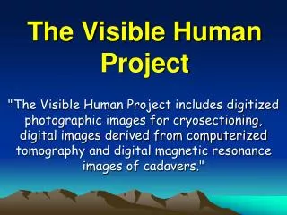
The Visible Human Project "The Visible Human Project includes digitized photographic images for cryosectioning, digital images derived from computerized tomography and digital magnetic resonance images of cadavers."
The Visible Human Project Find a normal male and female cadaver Fresh CT & MR Fill blood vessels and cavities with contrast media Pose and stabilize in opaque gel Freeze the cadaver Cut cadaver into blocks Frozen CT Grind down block and take photograph
Infuse blood vessels and fill cavities with contrast media (resin)
Data Collection CT (bone) MRI (proton-density) MRI (T1) MRI (T2)
Data Collection • Photographic cross-sections: Volume Dimensions: 1760 x 1024 x 1878Pixel Dimensions: .33 mm x .33 mm x 1 mmPixel Depth: 24-bit (8-bits x RGB)Volume Size: 9.5 GB • Computed Tomography (CT) - in fresh state: Volume Dimensions: 512 x 512 x 500Pixel Dimensions: variable x variable x 1 mmPixel Depth: 16-bitVolume Size: 250 MB • Computed Tomography (CT) – in frozen state: Volume Dimensions: 512 x 512 x 1878Pixel Dimensions: variable x variable x 1 mmPixel Depth: 16-bitVolume Size: 939 MB
Data Collection • Photographic cross-sections: Volume Dimensions: 1664 x 928 x 5189 Pixel Dimensions: .33 mm x .33 mm x .33 mm Pixel Depth: 24-bit (8-bits x RGB) Volume Size: 22.4 GB • Computed Tomography (CT) - in fresh state: Volume Dimensions: 512 x 512 x 1730 Pixel Dimensions: variable x variable x 1 mm Pixel Depth: 16-bit Volume Size: 864 MB
The Visible Human Transverse Coronal Sagittal
Semi-automatic Segmentation Mostly done by anatomists using a drawing and filtering program. Marr-Hildreth edge detection filter, seed filling and neighbor connectivity filter Spline interpolation of surface