
Uploaded by
bran
1 SLIDES
110 VUES
10LIKES
Immunohistochemical Detection of ASS in Melanoma Tissue
DESCRIPTION
Explore the expression of ASS in melanoma tissue through immunohistochemical analysis. A: Positive ASS staining shown in brown color. B: Melanoma sample lacking ASS expression. C: ASS-positive endothelial cells amidst ASS-negative melanoma tissue. Magnification: 200x.
Download
1 / 1
Télécharger la présentation 
Immunohistochemical Detection of ASS in Melanoma Tissue
An Image/Link below is provided (as is) to download presentation
Download Policy: Content on the Website is provided to you AS IS for your information and personal use and may not be sold / licensed / shared on other websites without getting consent from its author.
Content is provided to you AS IS for your information and personal use only.
Download presentation by click this link.
While downloading, if for some reason you are not able to download a presentation, the publisher may have deleted the file from their server.
During download, if you can't get a presentation, the file might be deleted by the publisher.
E N D
Presentation Transcript
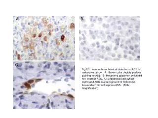
A B C Fig 2S. Immunohistochemical detection of ASS in melanoma tissue . A: Brown color depicts positive staining for ASS. B: Melanoma specimen which did not express ASS. C: Endothelial cells which expressed ASS in a background of melanoma tissue which did not express ASS. (200x magnification)
More Related