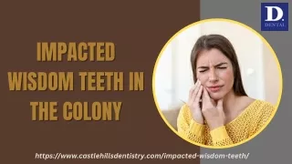
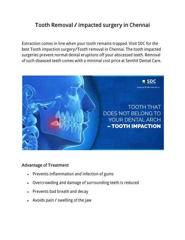
IMPACTED TEETH
Explore theories, causes, frequency, classification, and removal rationale of impacted teeth. Learn about surgical anatomy, postoperative care, and common complications. Discover the glossary of terms related to impacted and unerupted teeth.

IMPACTED TEETH
E N D
Presentation Transcript
Contents • Glossary of Terms • Theories of Impaction • Causes • Frequency • Classification • Rationale for Removal • Clinical & Radiological Assessment • Surgical Anatomy • Removal • Postoperative Care & Complications
Glossary of terms: • Impacted teeth: A tooth that has failed to erupt into normal functional position beyond the time usually expected for such appearance is impacted. Eruption is prevented by adjacent hard or soft tissue including overlying teeth, bone, or dense soft tissue.
Partial impaction: A tooth that is incompletely erupted is a partial impaction may be seen clinically but is frequently malposed and always covered to some extent with soft tissue or bone.
Unerupted teeth: A tooth that has not established normal full communication with the external environment of the oral cavity and remains contained within the hard or soft tissues of the jaw is referred to as Unerupted teeth.
Theories of Impaction By Durbeck 1) Orthodontic theory : Jaws develop in downward and forward direction. Growth of the jaw and movement of teeth occurs in forward direction any thing that interfere with such moment will cause an impaction (small jaw-decreased space). A dense bone decreases the movement of the teeth in forward direction. Causes for increased density of bone a) Acute infection, b) Local inflammation of PDL c) Malocclusion, d) trauma, e) Early loss of primary teeth – arrested growth of the jaw.
2) Phylogenic theory: Nature tries to eliminate the disused organs i.e., used makes the organ develop better, disuse causes slow regression of organ. [More-functional masticatory force – better the development of the jaw] Due to changing nutritional habits of our civilization have practically eliminated needs for large powerful jaws, thus, over centuries the mandible and maxilla decreased in size leaving insufficient room for third molars.
3) Mendelian theory: Heredity is most common cause. The hereditary transmission of small jaws and large teeth from parents to siblings. This may be important etiological factor in the occurrence of impaction. 4) Pathological theory: Chronic infections affecting an individual may bring the condensation of osseous tissue further preventing the growth and development of the jaws. 5) Endocrinal theory: Increase or decrease in growth hormone secretion may affect the size of the jaws
Causes of Impaction Local causes: • Irregularity in the position and pressure of an adjacent tooth. • The density of overlying or surrounding bone. • Long continued chronic inflammation with the resultant increase in density of the overlying mucous membrane. • Lack of space due to under develop jaws. • Prolong retention of the primary teeth. • Premature loss of primary teeth. • Acquired diseases – such as Necrosis due to infection or abscess and inflammatory changes in the bone due to exanthematous diseases in child.
Systemic causes: a) Prenatal causes – Hereditary Misagenation b) Post natal causes – All the conditions that may interfere with development of child. - Ricketts - Anaemia - Congenital syphilis - Tuberculosis - Endocrinal disfunction c) Rare conditions - Cleidocranial dysostosis - Oxycephaly - Progeria - Achondroplasia - Cleft palate
Frequency of impaction in following order • Mandibular third molars • Maxillary third molars • Maxillary cuspids • Mandibular bicuspids • Mandibular cuspids • Maxillary bicuspids • Maxillary central incisors • Maxillary lateral incisors
Classification Angulation – George Winter (1926) described first classification system which is based on the angulation of the long axis of the impacted third molar with respect to the long axis of the second molar. Mesioangular Horizontal
Vertical Distoangular
Buccal direction – Bucco version In addition impacted teeth also can be angled in buccal and lingual direction. Lingual direction – Lingual version Unusual position – torsiversion
Relationship to the anterior border of the ramus of the mandibular. Another method of classifying impacted third molar is based on the amount of impacted teeth that is covered with the bone of the mandibular ramus. [by Pell & Gregory] Class III: Class I : Class II
Relative depth of the third molar (Vertical plane) by Pell and Geogory : In this classification the degree is measured by the thickness of overlying bone, the degree of difficulty increases as the relative depth of third molar increases. Position A: Position B: Position C:
Killey & Key’s classification a) Based on angulation and position: Same as George Winters. b) Based on the state of eruption: - Completely erupted - Partially erupted - Unerupted c) Based on pattern of roots: 1) - Fused roots. - Two roots. - Two roots and multiple roots 2) Root pattern may be – - Surgical favourable - Surgical unfavourable
ADA classification: - Soft tissue impaction - Partial bony impaction - Complete bony impaction - Complete bony impaction with unusual complications
Rationale for removing impacted tooth.by Larry J. Peterson (JADA/Vol 123/1992 July) Indications: • Preventing and treating pericoronitis. • For prevention of dental caries. • Orthodontic considerations. • To prevent pathosis. • Prevention of root resorption. • Impacted teeth and dental prosthesis. • Prevention of dental diseases.
Contraindications: • Extremes of age. • Medically compromised patient. • Probable excessive damage to the adjacent structures. • Prevention of fracture of jaws.
Clinical Examination History: • Most patients are symptomatic. • If so then associated with- • (Pericoronitis / pain / swelling of the face / trismus / enlarged tender lymph nodes) Intraoral examination- • Size of oral cavity. • Degree of mouth opening. • Size of tongue. • Palpation for external oblique & internal oblique ridge in relation with 3rd molar.
Widely used radiographs: Periapical / OPG / Occlusion. Radiological assessment: • Orientation of the tooth. Position and depth of the tooth Winter lines.
As a general rule any tooth with redline 5mm or more is better remove under GA. If redline is 9mm or more in length the inferior surface of crown of impacted 3rd molar may be in level or even below the apex of 2nd molar White line: Red line: Amber line
Root pattern: Either Favourable Unfavourable • Shape of the crown. • Texture of investing bone. • Position and root pattern of 2nd molar. • Relationship of 3rd molar to the inferior dental canal.
Darkening of roots Deflection of roots Narrowing of roots Interruption of white line of canal Dark & Bifid apex Narrowing of canal Diversion of canal
A) Spatial Relationship Value - Mesioangular 1 - Horizontal / transverse 2 - Vertical 3 - Distoangular 4 B) Depth - Level A 1 - Level B 2 - Level C 3 C) Ramus relationship - Class I 1 - Class II 2 - Class III 3 DIFFICULTY INDEX Classification: Difficulty scores: Very difficult 7-10 Moderately 5-7 Minimally 3-4 Example: Mesioangular tooth 1 difficulty score is Level B 2 5-7 Class III 3 Moderately difficult
Temporalis muscle Buccinator Retro molar foramina SURGICAL ANATOMY OF MANDIBULAR 3RD MOLAR
Surgical Removal Following Steps: • Anaesthesia • Incision and mucoperiosteal flap. • Removal of bone. • Tooth removal. • Wound debridement. • Arrest of haemorrhage. • Wound closure. • Post operative followup.
Armamentarium (i) Local anesthesia (vi) needle holder (xi) cross bars (ii) 15 no. blade (vii) suture material (xii) retractors (iii) Tweezers (viii) scissors (iv) Curette (ix) chisels (v) Elevators (x) mallet
Various incisions / Approaches • Standard Wards incision. • Modified Wards incision. • Envelope flap. • L-Shaped flap. • Comma incision.
Ward’s incision: Modified Ward’s incision:
Envelop incision: L-shaped flap:
REMOVAL OF OVERLYING BONE I. Lingual split bone tech (Sir William Kelsey Fry) Advantages: • Quick & clean • Reduces the size of blood clot by means of saucerization of socket. Disadvantages: • Only suitable for young adults therefore Elastic Bone. • More chances of getting post operating lingual nerve parasthesia. • Patients inconvenience.
II. Moor / Gillbes Collor tech: • Conventional tech of using bur. • Similar amount of bone is sacrificed same as split bone technique. • Can be used in old patient with. • Convenient for patient. • Is to create a gutter along buccal side & distal surface of tooth. • And a point of elevation is created with bur.
III. Lateral Trephination tech: (Bowdler Henry) • Employed to remove any partially formed unerupted third molar that has not breached the hard & soft tissues overlying it. Advantages: Bone healing is excellent and here is no loss of alveolar bone around 2nd molar.
Sectioning TECHNIQUE The tooth is sectioned in different ways.
Horizontal impaction The tooth is sectioned in different ways.
Delivery of sectioned tooth from socket By using appropriate elevators. Straight elevator Warwick James Straight / Curved Coupland’s Cryer’s Cross bar Excessive force should be avoided to prevent injury Soft tissues Adjacent tooth / bone inferior dental canal / lingual nerve
Debridement of wound & closure • Thorough debridement of the socket by Periapical curette • Smoothening of sharp bony margins by Bone file / round burs • Thorough irrigation of the socket Betadine solution / Saline • Initial wound closure is achieved by Just distal to 2nd molar Posterior relieving incision Inter dental area mesial to 2nd molar 3-4 are usually sufficient
Post-operative care • Pressure pack • Ice pack • Avoid gargling / spitting • Soft diet • Warm water saline gargling after 12 hrs • Maintain oral hygiene • Proper medication
Complications • Soft tissue injuries : • facial vessels • Soft tissues • Lingual nerve • Inferior dental nerve • Bleeding – bone / soft tissue Intra-operative • Hard tissue injuries : • Osseous structures • Fracture of mandible • Injury to adjacent tooth
Post-operative • Immediate • Pain • Hemorrhage • Swelling • Trismus • Parasthesia • Late • Infection • Dry socket • Osteomylelitis • Secondary Hemorrhage • Pain in TMJ
Recent advancesUse of Erbium (Er):YAG laser [by M.Abu-Serriah / A.Ayoub : Bjoms 2004; 42: 203-208] Adv: • Less stressful • Less unpleasant • No vibrations & sound • Sharp clean cut through the bone & tooth • Can used anxious patients Disadv: • Compensate for tactile feedback compare to bur. • Trismus is more • Time consuming • Costly


![[PDF] Orthodontic and Surgical Management of Impacted Teeth Full](https://cdn7.slideserve.com/12521353/slide1-dt.jpg)