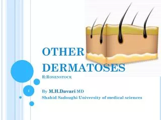
Dermatoses Resulting from Physical Factors
1.22k likes | 2.45k Vues
Dermatoses Resulting from Physical Factors. Chapter 3 Andrew’s Diseases of the Skin Adam Wray, D.O. November 15, 2005. Heat Injuries. Thermal Burns Electrical Burns Miliaria Miliaria Crystalline (Sudamina) Miliaria Rubra (Prickly Heat, Heat Rash) Miliaria Pustulosa Miliaria Profunda

Dermatoses Resulting from Physical Factors
E N D
Presentation Transcript
Dermatoses Resulting from Physical Factors Chapter 3 Andrew’s Diseases of the Skin Adam Wray, D.O. November 15, 2005
Heat Injuries • Thermal Burns • Electrical Burns • Miliaria • Miliaria Crystalline (Sudamina) • Miliaria Rubra (Prickly Heat, Heat Rash) • Miliaria Pustulosa • Miliaria Profunda • Occlusion Miliaria
Thermal Burns • First-degree burn- active congestion of superficial blood vessels • This causes erythema sometimes followed by epidermal desquamation • Constitutional reactions occur if area is large • Pain and increased surface heat may be severe
Deep Pale and anesthetic Injury to reticular dermis compromises blood flow and destroys appendages Healing takes > 1 month Scarring occurs Superficial Transudation of serum causing edema of superficial tissues Vesicles and blebs Complete recovery without scar or blemish is usual Second-degree burns
Thermal burn: This superficial second degree burn is characterized by bullae that contain serous fluid Second-degree burns
Second-Degree Burns • Inflicted scalds: severe second degree burns after dipping • B: two days after incident-to lower extremities and perineum • C: foot and lower leg
Second-Degree Burn • Accidental scald • Splash-and-droplet pattern of an accidental scald from hot cup of tea
Second-Degree Burn • Curling iron burn
Full-thickness tissue loss Skin appendages are destroyed There is no epithelium for regeneration Healing leaves a scar Third-degree burns
Fourth-degree burns • Destruction of entire skin and subcutaneous fat with any underlying tendons
Rule of nines: • In adults, an estimate of burn extent based upon this surface area distribution chart. Infants & children have a relatively increased head; trunk surface area ratio
Electrical Burns • Contact- small but deep, causing some necrosis of underlying tissues • Flash-burns usually cover a large area and are similar to a surface burn and should be tx as such • Lightening is the most lethal type of strike, cardiac arrest or other internal injuries may occur
Electrical Burns • Indirect- burns that are either linear in areas at which sweat was present; are feathery or aborescent pattern, which is believed to be pathognomonic
Electrical Burn • It is characterized by erythema, edema, bulla formation and sloughing of the necrotic epidermis
Electrical Burn-pathology • Blistering and elongated keratinocytes
Miliaria • Retention of sweat as a result of occlusion • Common in hot, humid climates • Occlusion of eccrine sweat gland obstructs delivery of sweat to the skin surface • Eventually backed-up pressure causes rupture of sweat gland or duct at different levels • Escape of sweat into adjacent tissue produces miliaria • Different forms of miliaria occur depending on the level of injury to the sweat gland
Miliaria Crystalline • Small, clear, superficial vesicles without inflammation • Appears in bedridden pts and bundled children • Lesions are asymptomatic and rupture at the slightest trauma • Self-limited; no tx is required
Miliaria Crystallina • Minute, discrete vesicles resulting from profuse sweating secondary to a high fever
Miliaria Rubra • Discrete, extremely pruritic, erythematous papulovesicles with sensation of prickling, burning, or tingling • Site of injury is prickle cell layer where spongiosis is produced
Miliaria Pustulosa • Always preceded by some injury, destruction, or blocking of sweat duct • Pustules independent of hair follicle • Seen in intertriginous areas, flexure surfaces of extrmities, sctrotum, and back of bedridden pts • Sterile pustules
Miliaria Profunda • Nonpruritic, flesh-colored, deep-seated, whitish papules • Asymptomatic, usually lasting only 1 hr after overheating has ended • Concentrated on the trunk and extremities • Occlusion is in upper dermis • Only seen in tropics usually following a severe bout of miliaria rubra
Occlusion Miliaria • May be produced with accompanying anhidrosis and increased heat stress susceptibility after application of extensive polyethylene film occlusion for > 48 hrs • Tx-place pt in a cool environment • Even a night in an air-conditioned room helps alleviate the discomfort
Occlusion Miliaria • Mild cases may respond to dusting powders, such as cornstarch or baby talcum powder • A lotion containing 1% menthol and glycerin and 4% salicylic acid in 95% alcohol is effective • An oily “shake” lotion such as calamine lotion, with 1% or 2% phenol may be effective
Erythema (pigmentatio) Ab Igne • Aka “toasted skin” syndrome • Persistent erythema or coarsely reticulated residual pigmentation resulting from it • Produced by long-continued exposure to excessive heat without production of a burn • It begins as a mottling caused by local hemostasis and becomes a reticulated erythema, leaving pigmentation
Erythema Ab Igne • Reticulated hyperpigmentation with some epidermal atrophy and scaling secondary to use of a heating pad
Use of bland emollients is helpful No effective treatment Kligman’s combination of 5% hydroquinone in hydrophilic ointment containing 0.1% retinoic acid and 0.1% dexamethasone may reduce unsightly pigmentation Histologically, an increased amount of elastic tissue in the dermis is seen Changes are similar to actinic elastosis, and has been suggested to call these changes thermal elastosis Erythema ag igne
Cold Injuries • Chilblain • Frostbite • Immersion injury
Chilblains • Acute chilblains is the mildest form of cold injury • Pts are usually unaware of injury until they develop burning, itching, and redness
Treatment • Nifedipine 20mg TID • Vasodilators (nicotina • amide 100 mg TID or dipyridamole 25 mg TID) • Systemic corticoid tx is helpful in chilblain lupus erythematosus • Pentoxifylline may be useful • Smoking strongly discouraged
When soft tissue is frozen and locally deprived of blood supply Frozen part is painless and becomes pale and waxy Four stages: I- Frost-nip erythema, edema,cutaneous anesthesia & transient pain II- second degree: hyperemia, edema & blistering, with clear fluid in bullae III- third-degree: full-thickness dermal loss with hemorrhagic bullae formation or waxy, dry, mummified skin IV- full-thickness loss of entire part Frostbite
Immersion Foot Syndromes • Trench Foot • Warm Water Immersion Foot
Trench Foot • Results from prolonged exposure to cold, wet conditions without immersion or actual freezing • Term derived from trench warfare in World War 1, when soldiers stood, sometimes for hours, in trenches with a few inches of cold water in them • Tx-removal from causal environment
Tropical immersion Foot • Seen after continuous immersion of the feet in water or mud of temperatures above 71.6 degrees F (22 degrees C) for 2-10 days • AKA “paddy foot” in Vietnam • Erythema, edema, and pain of the dorsal feet • Also fever and adenopathy • Resolution occurs 3 to 7 days after the feet have been dried
Dermatoses with Cold Hypersensitivity • Erythrocyanosis Crurum • Acrocyanosis • Cold Panniculitis
Slight swelling and a bluish pink tint of the skin of the legs and thighs of young girls and women May be unilateral May have cramps in the legs at night Small tender nodules may be found on palpation Nodules may break down and form small, multiple ulcers Seen in northern countries and probably due to an abnormal reaction of blood vessels to prolonged cold Erythrocyanosis Crurum
Acrocyanosis • A persistent cyanosis with coldness and hyperhidrosis of hands and feet • Chiefly occurs in young women • At times, on cold exposure, a digit becomes stark white and insensitive (acroasphyxia) • Cyanosis increases as the temperature decreases and changes to erythema with elevation of dependent part • Cause is unknown • Smoking, coffee, and tea should be avoided
Cold Panniculitis • After exposure to severe cold, well-demarcated erythematous warm plaques may develop, particularly on the cheeks of young children • Lesions usually develop within a few days after exposure, and resolve spontaneously in 2 weeks(approx) • No tx is indicated • Popsicle dermatitis is a temporary redness and induration of the cheek in children resulting from sucking Popsicles
Parts of solar spectrum important to photomedicine: Visible light 400 to 760 nm Infrared radiation beyond 760 nm Visible light has little biologic activity, except for stimulating the retina Infrared radiation is experienced as radiant heat Below 400 nm is the ultraviolet spectrum, divided into three bands: UVA, 320 to 400 nm UVB, 290 to 320 nm UVC, 200 to 290 nm Virtually no UVC reaches the earth’s surface, because it is absorbed by the ozone layer Exception: Australia, welders Sunburn and Solar Erythema
UVB is 1000 times more erythemogenic than UVA UVA is 100 times greater than UVB radiation during the midday hours Most solar erythema is cause by UVB Sunlight early and late in the day contains more UVA UVA is reflected from sand, snow, or ice to a greater degree than UVB Amount of ultraviolet exposure increases at higher altitudes, is greater in tropical regions, and temperate climates in summer Sunburn and Solar Erythema
Clinical signs and symptoms • Sunburn is normal cutaneous reaction to sunlight in excess of an erythema dose (the amount that will induce reddening) • UVB erythema peaks at 12 to 24 hrs after exposure • Desquamation is common about a week after sunburn even in non-blistering areas
Sunburn treatment • Cool compresses • Topical steroids • Topical remedy: Indomethacin 100 mg Absolute ethanol 57 ml Propylene glycol 57 ml spread widely over burned area with palms and let dry
Prophylaxis • Avoid sun exposure between 10 am and 2 pm • Barrier protection with hats and clothing • Suncreen agents include UV-absorbing chemicals and UV-scattering or –blocking agents(physical sunscreens)
Chemical suncreens-para-aminobenzoic acid(PABA), PABA esters, cinnamates,salicylates, anthranilates, benzophenoes) Physical agents-titanium dioxide Combinations of the two Water resistant-maintaining their SPF after 40 minutes of water immersion Water proof-maintaining their SPF after 80 mins of water immersion UVA protection- sunscreens containing benzophenones or dibenzoylmethanes Apply sunscreen at least 20mins before sun exposure Sunscreens