PANCREATITIS
370 likes | 590 Vues
PANCREATITIS. SAI YAN AU CHEE SIONG KOH. INTRODUCTION. Mrs Emilie Rezek, a 78 years old elderly female, presents with an hour’s history of severe epigastric pain radiating to the back. PAST HISTORY. “ Bad turn” of the heart about 1 month ago Admitted to RHH and had a cardiac stenting

PANCREATITIS
E N D
Presentation Transcript
PANCREATITIS SAI YAN AU CHEE SIONG KOH
INTRODUCTION • Mrs Emilie Rezek, a 78 years old elderly female, presents with an hour’s history of severe epigastric pain radiating to the back.
PAST HISTORY • “Bad turn” of the heart about 1 month ago • Admitted to RHH and had a cardiac stenting • On a background of chest pain 6 to 7 years ago • Exertion induced • On medication – Loprex • Worsening over the 6 to 7 years • Acute Myocardial Infarction 10 years ago
Long standing stomachache (DYSPEPSIA) – gastroscopy in 1994 • Cholecystectomy • On Zoloft
No hypertension • No diabetes mellitus • No asthma and epilepsy • NKA
PRESENTING COMPLAINT(PC) • EPIGASTRIC PAIN
HISTORY OF PC • EPIGASTRIC PAIN • Started 1 day after discharge from RHH • Was sleeping at time of attack • Lasted for 15 min and spontaneous resolve for 10 min and started again – call for ambulance • Ripping pain on a scale of 6/10(1st episode) and 10/10( 2nd episode) which radiates to the back
EPIGASTRIC PAIN • No relieving or aggravating factors • Other associated features includes shortness of breath, nausea and perspiration(diaphoresis)
FAMILY HISTORY • Father died in world war II – traumatic injuries • Mother died in world war II – marrow problem • Has 3 sons: • One has heart disease • One died of traumatic injuries • One is a/w
SOCIAL HISTORY • Came from Prague in 1949 with husband • Lives alone since death of husband • Able to cook and clean the house until 2 months ago when shortness of breath worsened • Non smoker • Non drinker • Has a dog
SYSTEMIC REVIEW • Cardiovascular system • Chest pain • Shortness of breath of few years worsened 2 to 3 months ago • No palpitation • No PND
Gastrointestinal • Vomiting for 3 to 4 times for past 2 months • Constipated for past 2 months – goes to toilet every 3 days • No loss of appetite • No loss of weight • No diarrhoea • Good energy status
Cardiovascular • Chest pain • Shortness of breath worsened 2 to 3 months ago • No palpitation • No PND
Other systems • No positive findings
CLINICAL EXAMINATION • Elderly lady • Not cyanosed or jaundiced • Non cachectic • Afebrile • Heart rate - 76/min, regular rhythm • Blood pressure - 140/80 mmHg • Respiratory rate - 20/min
Positive findings : - • GIT • Right cholecystectomy scar • Right upper quadrant tenderness • Non distended abdomen • No organ enlargement • No enlarged lymph nodes • No palpable mass • No ascites • No renal bruit • Bowel sound presence
CVS, RESPI, MSK, GUT, HAEMO, ENDO AND NEURO – no abnormal findings
DIFFERENTIAL DIAGNOSIS • Pancreatitis • Peptic ulcer disease ? • Acute Myocardial Infarction ?
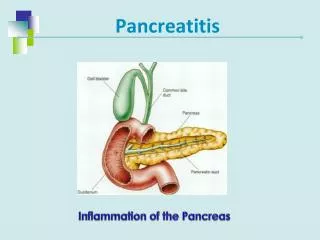
LOCATION OF PANCREAS • Elongated structure that lies in the epigastrium and the left upper quadrant. • Soft, lobulated and situated on the posterior abdominal wall behind the peritoneum. • Crosses the transpyloric plane • Divided into a head, neck, body, and tail.
THE PANCREAS • Here is the normal gross appearance of the adult pancreas; a small portion of duodenum is at the left next to the head; the tail of the pancreas is at the right. The pancreas has a tan, lobular architecture. Adjacent adipose tissue and lymph nodes are closely apposed.
PANCREATITIS • Definition : - • An inflammatory disorder of the pancreas • Characterized by abdominal pain • Attacks range from mild to severe • Can occur as acute form (acute pancreatitis), recurrent acute form (chronic relapsing pancreatitis) and persistent (chronic pancreatitis)
ACUTE PANCREATITIS • Mild, self -limited, and more serious • Female > male • Peak incidence - between 50 to 60 YO • Most common causes - gallstones, alcohol • Other causes - iatrogenic or traumatic, metabolic, infection and idiopathic
Pathophysiology : - • Sudden onset of diffuse inflammation • Acute haemorrhage • Causes extensive necrosis of the organ • Clinical features : - • Mild attack • Moderate attack • Severe attack
This is an example of acute pancreatitis. The pancreas is swollen and does not show the typical tan, lobulated architecture. Instead, it has areas of hemorrhagic necrosis that appear as blotchy black red areas at the mid right of the photograph.
Mild attack • Acute abdominal pain • Minimal/rapidly resolving abdo signs • Minimum systemic illness • Moderate attack • Severe acute abdo pain • Tachycardia • Abdo distension, tenderness and guarding
Severe attacks • Severe acute abdominal pain • Severe toxaemia and shock • Generalised peritonitis • ARDS Reference : Essential Surgery, Burkitt,Quick and Gatt, 2nd Edition, page 275
INVESTIGATIONS • Serum amylase level ( >1200 i.u/mL) • Liver Function Tests (ALT) – raised 3 times more than normal in gallstone pancreatitis • Plain x – rays of chest ( erect ) and abdomen ( supine ) • ERCP • Peritoneal tap
MANAGEMENT • “NIL by mouth” and I/V fluids • Nasogastric aspiration • Broad - spectrum I/V antibiotics • Intensive care • Fluild and electrolyte management • Treatment of hypocalcaemia • Ventilatory support • surgery
CHRONIC PANCREATITIS • Continuous or relapsing inflammation of the pancreas leading to irreversible morphologic damage and permanent impairment of function • Etiology – alcohol, gallstones • Pathophysiology : - • Loss of pancreatic parenchyma • Replacement by fibrous tissue
Clinical features : - • Persistent and severe upper abdominal pain • Steatorrhoea, vomiting, abdo distension and progressive weight loss.
INVESTIGATION • Serum amylase • Ultrasound • Abdominal x-ray • CT scan • ERCP
MANAGEMENT • Medical intervention • Treat the pain • Maintain nutritional status • Reduced symptoms
Surgery • Removal of pancreatic duct stones • Partial pancreatectomy of the head and tail • Sphincteroplasty of the pancreatic duct opening