
Selective Self-Assembly of Biogenic Silica via Layer-by-Layer Deposition and Inkjet Printing
This research explores the selective self-assembly of biogenic silica derived from diatom shells using Layer-by-Layer (LbL) deposition and inkjet printing. Diatom frustules, with intricate nanoscale features, are patterned on polyelectrolyte layers, facilitating the creation of luminescent materials and photonic structures. The method enhances the potential of diatom structures in sensor applications, leveraging their unique optical properties. The study showcases the fabrication of a 2D diatom shell matrix and highlights future applications in gas sensors and photonic crystals.

Selective Self-Assembly of Biogenic Silica via Layer-by-Layer Deposition and Inkjet Printing
E N D
Presentation Transcript
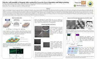
Selective self-assembly of biogenic silica assisted by Layer-by-Layer deposition and Inkjet printing Wei Wang, Doo-HyoungLee, Clayton Jeffryes, Debra Gale, Gregory L. Rorrer, Timothy Gutu, Jun Jiao, and Chih-hung Chang Oregon State University, Department of Chemical Engineering, Corvallis, OR 97330 Portland State University, Department of Physics, Portland, OR 97201 Diatom shells - - Electrostatic force interaction + + - - + + Positively charged polyelectrolyte layer Multilayer film Abstract Nature is an inspirational source of silica structures that possess unique optical properties. A few classes of organisms fabricate metal oxides with nanoscale features by a bottom-up self assembly process. In particular, diatoms are a prolific class of single-celled algae that possess silica shells or “frustules” with intricate submicron scale features, including two-dimensional pore arrays. During frustule development, membrane-bound transporters actively take actively up the soluble silicon in the form of Si(OH)4. Once inside the cell, Si(OH)4 is converted to nanostructured silica by protein-mediated condensation reaction within the silicon deposition vesicle, an organelle which serves as the mold for frustule development. Recently, it has been shown that intact diatom frustules can act as 2D photonic crystals and optical sensor platforms for the detection of organic vapors. Techniques to assemble and pattern these microorganisms at large scale is needed to take advantage of these nanostructured silica for device applications such as sensors, photonic crystals and electroluminescence devices, Herein, biogenic silica fabricated from marine diatom whose cell walls possess intricate nano- and microstructures was selectively adsorbed on polyelectrolyte layers which was patterned by inkjet printing. In addition, we will also describe a new technique to fabricate patterned nanostructured silicate-based luminescent materials by inkjet printing. Substrate Experimental procedure Biogenic silica based phosphor materials patterned by inkjet printing Doped with Rhodamine-123, printed PAH layers can be visualized in fluorescence images. Based on intensity of green, one can observe that the PAH films are uniform across the printed area. The size of PAH dots is about 250µm which is similar to the dimension of cosco sp. diatom frustules. 2-Dimensional diatom shell matrix After deposition diatom silica shells (cosco sp. ~200µm in diameter) were arranged in a 2-dimentsion matrix with preservation of submicron structure. Only one diatom shell seats on a PAH dot controlled by the area of printed PAH dots. TEM images and EDX analysis on frustules show the crystalline of Y2SiO5:Eu3+ which was formed by printing yttrium precursor solution on deposited frustule layers. Using Layer-by-Layer deposition, positively charged Poly(sodium 4-stryrene sulfonate) (PSS) and negatively charged Poly(allylamine hydrochloride) (PAH) were alternatively deposited on glass followed by inkjet-printing a PAH layer which attracted frustules on them, resulting in a frustule-matrix. Mechanism Zn2SiO4:Mn2+ and Y2SiO5:Eu3+ had photoluminescence emission spectra in green and orange. The words ‘green’ and ‘OSU’ in the image are made from frustules and precursor inks via inkjet printing. Using smaller diatom shells (cyclotella sp. ~10µm ) uniform frustules monolayers were formed on printed PAH dots. Due to strong Brownian motion, Cyclotellafrustules are easier to pattern compared to larger species. OH groups on the diatom shells surface endow frustules with negative charges which are attracted by positive charges on PAH layer and expelled by negative charges on PSS via electrostatic force. That results in the selective adsorption of biogenic silica. Future work Sketch of Gas sensor matrix Idea sketch of gas sensor matrix with diatom shells which are selectively converted to sensing materials by inkjet- printing various precursor inks on each diatom shell. The large surface/volume ratio of frustules enables them to be a good candidate for the device of sensors. Printed PAH layer The adsorption of biogenic silica is controllable in aspect of patterning. One can adjust the assembly via varying the pH and ionic strength of suspension of frustules and also the area of printed PAH dots. The attractive point in this research is the potential application of 2-dimensional diatom shell matrix into gas sensor and 2-dimensional photonic crystals. Diatom sensors Conductive lines This research was supported by the National Science Foundation (NSF) under Nanoscale Interdisciplinary Research Team (NIRT) award number BES-0400648.


