Understanding Membrane Potential and Action Potential in Medical Physiology
360 likes | 1.87k Vues
Learn about the resting membrane potential, generation and propagation of action potentials, nerve conduction, and more in excitable tissues. Explore the electrochemical basis behind cell membrane function.

Understanding Membrane Potential and Action Potential in Medical Physiology
E N D
Presentation Transcript
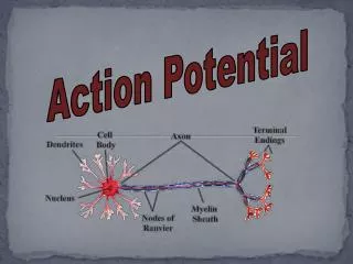
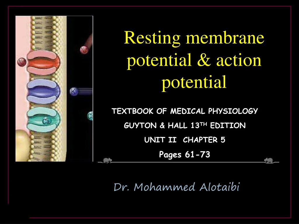
Resting membrane potential & action potential TEXTBOOK OF MEDICAL PHYSIOLOGY GUYTON & HALL 13TH EDITION UNIT II CHAPTER 5 Pages 61-73 TEXTBOOK OF MEDICAL PHYSIOLOGY GUYTON & HALL 13TH EDITION UNIT II CHAPTER 5 Pages 61-64 Dr. Mohammed Alotaibi
Objectives At the end of these lectures the student should be able to • Explain why some membranes areexcitable. • Describe the electrochemical basis ofRMP. • Describe the mechanism of generation and propagation ofAP. • Describe conduction along nerve fibers, role of myelination and how nerve fibers are classified.
ExcitableTissues • Tissues which are capable of generation and transmission of electrochemical impulses along themembrane Nerve Muscles
Excitabletissues excitable Non-excitable neuron Redcell GIT muscle •RBC •Intestinalcells •Fibroblasts •Adipocytes •Nerve •Muscle •Skeletal •Cardiac •Smooth
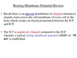
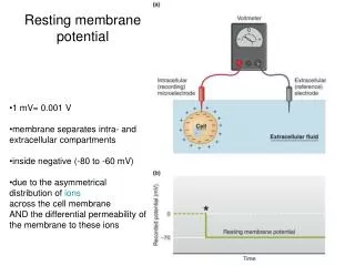
Membranepotential • A potential difference exists across all cell membranes • This iscalled • – Resting Membrane Potential(RMP)
Membranepotential • Inside is negative with respect to theoutside • This is measured using microelectrodes and oscilloscope (VOLTMETER) • This is about -70 to -90mV
Excitabletissues Non-excitable excitable neuron Redcell GIT muscle • Non-excitable tissues have less negativeRMP • -53 mV epithelialcells • -8.4 mVRBC • -20 to -30 mVfibroblasts • -58 mVadipocytes • Excitable tissues have more negativeRMP • ( - 70 mV to - 90mV)
Resting MembranePotential • This depends on the followingfactors • Ionic distribution across themembrane • Membranepermeability • Otherfactors • Na+/K+pump
Ionicdistribution Na+ Cl- K+ Pr- • Majorions • Extracellularions • Na+,Cl- • Intracellularions • K+,Proteins
Ionicchannels Na+ Out In K+ • Leaky channels (K+/Na+leakchannels) • More permeable toK • Allows free flow ofions • In the restingstate • K+ permeability is 100 times more than that ofNa+
Na+/K+pump 2K+ ATP 3Na+ ADP • Active transport system for Na+-K+ exchange usingenergy • It is an electrogenic pump since 3 Na+ efflux coupled with 2 K+influx • Net effect of causing negative charge inside the membrane
Factors contributing toRMP • One of the main factors is K+ efflux (NernstPotential:-94mV) • Contribution of Na+influx is little (NernstPotential:+61mV) • Na+/K+pump creates additional degree of negativity inside themembrane (-4mV) • Negatively charged protein ions remaining insidethe membrane contributes to thenegativity • Net result: -70 to -90 mVinside
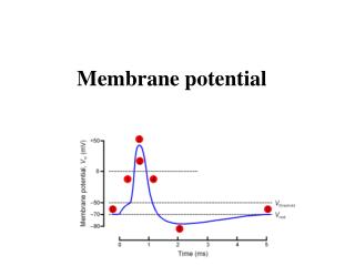
The action potential Nerve signals are transmitted by action potentials, which are rapid changes in the membrane potential that spread rapidly along the nerve fiber membrane to produce physiological effects such as: • Transmission of impulse along nerve fibres • Release of neurotransmitters • Muscle contraction • Activation or inhibition of glandular secretion
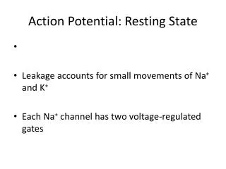
Each action potential begins with a sudden change from the normal resting negative membrane potential to a positive potential and ends with an almost equally rapid change back to the negative potential. Stages of the action potential: Resting Stage. It is the resting membrane potential before the action potential begins. The membrane is “polarized”. Depolarization Stage. Repolarization Stage.
Depolarization Na+ • Depolarization:The membrane suddenly becomes permeable to Na+ions, allowing tremendous numbers of positively charged Na+to diffuse to the interior of the axon (Upstroke).
Repolarization Na+ K+ Repolarization:Na+channels begin to close and the K+channels open. Rapid diffusion of K+ions to the exterior re-establishes the normal negative resting membrane potential.
Reversal Potential = + 35 mV Local Responses Threhold Potential ( Firing Level ) = -50 to -65 mV RMP= -90 mV Q : What opens the voltage-gated channels ? Opened by a stimulus strong enough to depolarize them to threshold Increasing Stimulation
Threshold stimulus: • The membrane potential at which occurrence of the action potential is inevitable. Acute subthreshold potential: • Stimulus that resultsonly in local depolarisation (acute local potentials) when stimulus is below the threshold. Local Responses All-or-nothing principle: Once threshold value for excitation is reached a full AP is produced, its intensity can not be increased by increasing stimulus intensity.
Types of transport channels through the nerve membrane: • Voltage gated Na+ channels • Voltage gated K+ channels
Voltage gated Na+ channels At rest, the activation gate is closed and the inactivation gate is open. During the upstroke of the action potential, both gates are open and Na+ flows into the cell down its electrochemical potential gradient. During repolarization, the activation gate remains open but the inactivation gate is closed. local anestheticlidocaine blocks this channel Cannot elicit new AP
The Na+ Voltage-Gated Channel (1) • Has 2 gates : one on the outer side of the membrane and is called the activation gate , • and another one on the inner side of membrane called the inactivation gate . • And this channel has 3 states : • (1) Resting state : in the resting cell , when the MP = RMP = -70 to -90 mV , • the activation gate is closed • this prevents entry of Na+ to the interior of the cell through this gate.
Activated State of Sodium Channel • (2) Activated state : when a Threshold Depolarizing Stimulus moves the MP from its resting value (-90 mV ) to its Threshold value (-65 to -55mV) • this opens the activation gate , and now the Na+ channel is said to be in the Activated State • ( NB in this case BOTH the activation gate & inactivation gate are open ) • permeability to Na+ becomes increased 500 to 5000 times Na+ influx • Na+ flows into the cell in large amounts ,
Inactivated State of Sodium Channel • (3) Inactivated state : A few milliseconds after the activation gate opens , the channel becomes inactivated : At the peak of AP the inactivation gate will close • the inactivation gate will not open by a second stimulus & the cell becomes Refractory ممانعة ) to another stimulation . • This goes on until the MP has gone back to its resting ( RMP) level ( -70 to -90mV). • in this case , while the activation gate is still open , • the inactivation gate is closed .
Voltage gated K+ channels • Has one gate only . • During the resting state, the gate of the potassium channel is closed and potassium ions are prevented from passing through this channel to the exterior. • Shortly after depolarization, when the sodium channel begins to be inactivated, the potassium channel opens. K+ exits (Efflux) Repolarization
Hyperpolarization: Why? • For a brief period following repolarization, the K+ conductance is higher than at rest. • Na +-K +ATPase pumpnow starts to move Na+ out & K+ in against their concentration gradient. Na/K Pump brings membrane potential back to its resting value Hyperpolarization
Refractory Periods Two stages • Absolute refractory period The period during which a second action potential cannot be elicited, even with a strong stimulus. • Relative refractory period Can trigger new action potential if stimulus is very strong. Higher K+ conductance than is present at rest Closure of the inactivation gates of the Na+ channel
Conduction Velocity It is the speed at which action potentials are conducted (propagated) along a nerve or muscle fiber. The larger the diameter, the faster the transmission, Because: -Large fiber offers Less resistance to local current flow & more ions will flow. Mechanisms that increase conduction velocity along a nerve: 1- Nerve diameter. Faster conduction Slower conduction
Conduction Velocity Mechanisms that increase conduction velocity along a nerve: 2- Myelination. Myelin is an insulator that makes it more difficult for charges to flow between intracellular and extracellular fluids. • The layers of Schwann cell membrane contain the lipid substance sphingomyelinwhich is excellent electrical insulator that decreases ion flow through the membrane. • - Node of Ranvier: small uninsulated area where ions can flow with ease.
Saltatory Conduction It is the jumping of action potentials from one node of ranvier to the next as they propagate along a myelinatedfiber. Value:- 1- Increases conduction velocity. 2- Conserves energy for axon because only nodes depolarize.
What happens if myelination is lost? • Multiple sclerosis • Autoimmune disease (Immune system attacks the myelin sheaths surrounding axons as well as the axons themselves). • Usually young adults • Blindness, problems controlling muscles • Ultimately paralysis • Scar tissues (scleroses) replaces some damaged cells.