Mitral Apparatus
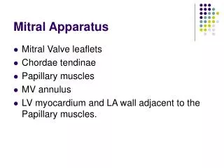
Mitral Apparatus. Mitral Valve leaflets Chordae tendinae Papillary muscles MV annulus LV myocardium and LA wall adjacent to the Papillary muscles. Incidence of Mitral Regurgitation. Framingham study found mild MR present in 19% of men and woman (asymptomatic)

Mitral Apparatus
E N D
Presentation Transcript
Mitral Apparatus • Mitral Valve leaflets • Chordae tendinae • Papillary muscles • MV annulus • LV myocardium and LA wall adjacent to the Papillary muscles.
Incidence of Mitral Regurgitation • Framingham study found mild MR present in 19% of men and woman (asymptomatic) • Severe MR, dependent on the study, is present from 0.2-1.9% of the general population.
Mitral Regurgitation • Acute primary MR. • Chronic primary MR. • 3. Secondary MR.
Ischemic papillary m. dysfunction • Usually occurs with acute MI • Can occur wth anterior or inferior MI • May occur with angina and then resolves • Papillary m. complete rupture has a very high mortality
Ischemic MR • About 10% of MI have MR by cath • Moderate to severe in 3-4 % • Some ECHO studies after MI show varying degrees of MR in +- 50 % of cases • Old patients and woman have a higher incidence.More common with large MI • Incidence ↓ with early reperfusion of MI • Surgery ( MVR ) may be indicated and has a high morbidity and mortality.
Causes of secondary MR • CAD, old MI, ischemic cardiomyopathy • Idiopathic dilated cardiomyopathy • Advanced aortic valve disease • Hypertensive heart disease • Any cardiac disease that leads to LV dilatation
Clues that MR is secondary • ECG = Severe LVH,MI. • Rhythm is NSR ( not a fib ) • LA is not disproportionately large • EF is low ( uncommon in primary MR ) • MV appears normal • Segmental wall motion abnormalities on Echo, MI on ECG
Common causes of acute primary MR • Endocarditis involving the valve leaflets, cordae tendineae or annulus. • Spontaneous ( idiopathic ) rupture of cordae • Rupture of diseased cordae ie. Collagen vascular disease , Marfanoid state, RHD or MV prolapse syndrome.
Common causes of Chronic primary MR • Any of the causes of acute primary MR may progress to chronic primary MR • Mitral prolapse syndrome • Chronic RHD
Examples of ECHO in MR • RHD. MV thick and some MS • Endocarditis = vegetations • Chordal rupture = ↑ leaflet motion • MVP often causes hammocking or posterior motion of leaflets. • Failure of leaflets to close completely suggests papillary m. dysfunction or a markedly ↑ LV size.
Pathophysiology of Chronic MR • Isovolumetric contraction phase absent • Afterload greatly ↓ • LV dilatation occurs 1st in flow loads • EF initially ↑ and remains high or normal • EF falls very late in disease • LA slowly dilates and may be enormous • CO well preserved • Symptoms may not occur for many years • Severe LVH does not occur
ECG IN CHRONIC PRIMARY MR • Atrial fibrillation is very common. • If in NSR P wave broadens to greater than 0.12 secs. • P wave be notched and have a large negative component in V1 • If pulmonary hypertension present, RVH, tall R in V1. • Mild LVH ( one should never see severe LVH if only Chronic MR present )..
Symptoms of MR • Symptoms are related to the decrease in forward effective cardiac output and LA pressure. • Weakness,fatigue and poor exercise tolerance • LV failure symptoms • R heart failure ( less common) • Thromboembolism
Physical Exam in chronic primary MR 1.Arterial pulse is ↓ in volume but is brisk (reflecting high EF). Pulse pressure usually is normal-not wide as in chronic AI. 2.S1 ↓ reflecting failure of leaflets to close properly. 3.S2 may be widely split due to ↓ in LV ejection time and delay of S2P if pulmonary hypertension is present. 4.Murmur is systolic but its duration, quality, location and radiation depend on the component of the Mitral apparatus effected. 5.In most case pansystolic murmur starts with S1 and continues to S2. It is heard best at apex and radiates to axilla or back, usually blowing and high pitched in quality.
ATYPICAL MR MURMURS 1.When posterior leaflet involved due to prolapse or chordal rupture, murmur may radiate to sternum or base of heart. 2.Anterior leaflet involvement in prolapse may be very loud and radiate to neck. 3.MV prolapse M may be harsh and be mid or late systolic,preceeded by a mid systolic click. 4.M of Pap.Muscle ischemia may be variable and become louder during an ischemic episode. 5.Large diastolic volumes across valve may produce a diastolic murmur. 6.Secondary MR murmur may decrease with RX of CHF. 7.No murmur,particularly if CO low
EFFECT OF DIAGNOSTIC MANEUVERS ON MR • MR may be louder when LV volume increases as in CHF or leg raising when supine. • 2. May increase when afterload increases ( squating or isometric handgrip ). • 3. May be softer when venous return is decreased as in standing or Valsalva. • 4. MVP murmur is longer when patient stands and click moves closer to S1.
Clinical manifestations of MR. • The nature and severity of MR symptoms depends on the: • Amount of MR • Rate of progression of MR • Acute or chronic • LA pressure(or Pa wedge pressure) • PA pressure • Associated cardiac disease
ECHOCARDIOGRAM IN MR. • With M mode and 2 D Echo (TTEorTEE) there is ↑ LA, • ↑ LV and LV contraction pattern ( EF ↑ ). • Mechanism of MR usually identified. • Prolapse,endocarditis,Flail leaflet,RHD etc. • Colorflow Doppler gives degree of MR • Degree of MR is just an estimate
Indications for TEE • When TTE is of poor quality • When endocarditis suspected and TTE is equivocal • When endocarditis is suspected and TTE is negative ( will sometimes see a vegetation with TEE ).
Cardiac Cath. In MR • Pressure recordings may show giant V waves in major acute MR. • Ventriculography done when Echo is poor and degree of MR uncertain • Coronary angiography when ischemic heart disease suspected. • Pressure data may be helpful to decide whether symptoms are due to MR.
Surgery in ACUTE MR • Torn chord and acute primary MR may cause intractable pulmonary edema requiring MVR • Papillary M. rupture in acute MI may require MVR if med RX fails. • Endocarditis with 4+ MR, surgery if patient can’t be stabilized medically.
Surgery in Chronic Primary MR • MR may progress insidiously without symptoms. • If CHF occurs, surgery indicated. • Surgery indicated in asymptomatic MR when ( 55 rule applies ) • End systolic dimension approached 55mm or EF falls below 55% (older criteria ). • Surgery not performed in secondary MR but may be considered if MR 4+ and EF above 30%.
Indications for surgery in asymptomatic chronic MR • Monitor LV size and EF • Surgery indicated when end systolic dimension approaches 55 mm ( now 40 mm ) • Surgery indicated when EF is falling and approaches 55% (now 60%) • Some newer recommendations are based on the amount of MR. ( NEJM 3/3/2005, VOl. 352:875-883 ) .Pts with effective regurgitant orifice > 40mm2 need surgery. • Catheter techniques for secondary MR ? Indications • New consideration,elevated BNP levels may help in deciding whether to operate
MV SURGERY IN CHRONIC SEVERE MR ( 2006 guidelines) in JACC VOL.48,no.3,2006 MV surgery is beneficial for asymptomatic patients with chronic severe MR and mild to moderate LV dysfunction, EF 0.30-60% and/or end-systolic dimension greater than or equal to 40mm. (level of evidence B)
Mitral Valve surgery • Mechanical valve ie St. Jude. Advantage: longevity. Problem: Warfarin • Tissue valve. Advantage: no Warfarin.Problem: longevity • MV repair. Advantage: better LV function , no Warfarin,problem: may not be technically feasible.
Indications for Surgery for MR • Preop LV function is best predictor of long term morbidity and mortality • Earlyl surgery for severe MR is key. • Recommendations for surgery • Any pt.with severe MR even if no symptoms,but good LV function,as long as • Low op risk and chance for valve repair • Asymptomatic patients with NL EF but effective regurgitant orifice by Doppler is 40 mm squared or more.
Functional anatomy and outcome of MR surgery • TTE and particularly TEE may be very helpful in deciding which valves can be repaired. • Patients with floppy valves are often good candidates for repair. • Very calcified valves, ischemic MR and secondary ( functional ) MR patients are usually not candidates for repair.
Indications for Surgery in MR • Consider waiting for signs of decreased LV FX or progressive dilatation in pts with: • Less than severe MR • High OP risk due to co-morbidities • Likelihood of repair low • Narcotic addicts with endocarditis