Download
1 / 29
360 likes | 2.38k Vues
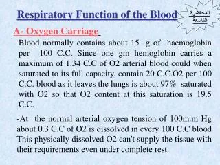
Function of blood and blood plasma. Dpt. of Normal, Pathological and Clinical Physiology Charles University, 3 rd Faculty of Medicine. The main function of blood. Respiration (transport of O 2 a CO ´2 ) Nutrition (transport of ingested nutrients) Transportation of waste products

E N D
Function of blood and blood plasma Dpt. of Normal, Pathological and Clinical Physiology Charles University, 3rd Faculty of Medicine
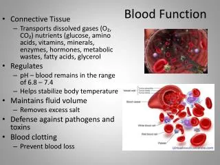
The main function of blood • Respiration (transport of O2 a CO´2) • Nutrition (transport of ingested nutrients) • Transportation of waste products • Transport of heat (for heating and cooling) • Acid-base balance • Water balance • Thermoregulation • Immunity • Transport of hormones (signals), vitamins and trace elements • Hemocoagulation (hemostasis)
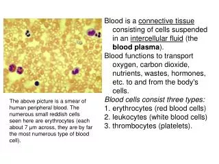
Main components • Whole blood (8% of body weight) =blood elements + blood plasma • erythrocytes 4.2 – 6.0 x10¹²/l • leukocytes 3 – 11 x109/l • thrombocytes 170 – 360 x109/l • serum x plasma • Hematokrit 36% - 49%
Composition of blood plasma • water • sodium 135-150 mmol/l, potassium 3.8-5.5 mmol/l, calcium 2.0-2.75 mmol/l, magnesium 0.66-0.94 mmol/l • chlorides 97-108 mmol/l, bicarbonate, phosphate, sulphate, proteins 70-80 g/l • glucose 3.3-6.1 mmol/l, urea 2-7.5 mmol/l • viscosity (water=1): blood 4.5, plasma 2.2 • osmolality: 280 mosm/l (the major cation is Na, the major anions are HCO3 and Cl
Plasma proteins • Oncotic pressure (colloidal-osmotic) (3 kPa), edema • Synthesis in liver • glycoprotein (except albumin) • Proteins of acute phase (CRP) • 70-80 g/l • Blood volume
1. Albumin • 32-45 g/l, 69 kDa, 60 % of all plasma proteins, 80 % of oncotic pressure • 12 g/day produced in liver (25 % of capacity) • Liver diseases – decrease of A:G ratio • 585 AK, ellipsoidal shape (15 x 3.8 nm) • albuminuria • transport: FFAcids, Ca, bilirubin, steroid hormones, Cu, penicillin, aspirin
2. Haptoglobin • Glycoprotein binding free Hb (10 % Hb of destroyed erythrocytes, the rest breaks down into globin, hem and iron), 0.4- 1.8 g/l of Hp, the same amount of Hb, 90 kDa • free Hb is filtrated in kidney and may affect tubules (transfusion) • Hp-Hb complex is not filtrated: iron sparing and tubules protecting effects • Decreases during hemolytic anemia (half-times of Hp-Hb and Hp), increases during inflammation (PAF)
3. Iron coupled proteins • transferin (2-4 g/l), feritin (plasma level corresponds to the body reserve), hemosiderin • hemochromatosis
4. Ceruloplazmin • 2-globulin, 160 kDa, 0.3 g/l • Transfer of 90 % of copper (6 atoms bind to one molecule), the rest is transported bound to albumin, easy release = probably more important) • Connected to the Wilson disease (hepatolenticular degeneration, AR, storage of copper in the brain, cornea, kidney and liver, high intestinal adsorption and low liver excretion of copper; Hepatitis, anemia, neurological signs, Kayser-Fleischer ring; )
5. 1-antitrypsin • The main component of 1 fraction • Inhibits the trypsin, elastase and other protease • deficiency (mutation) results in accumulation of the 1-AT in hepatocytes, hepatitis and cirrhosis (unknown mechanism), transplantation
6. Immunoglobulins • Produced by plasma cells (B-lymphocytes) • antibodies, the defense proteins
The most numerous cell of the human body (2.5x10¹³), the speed of production (2.5 mil./s), 4 kms daily diameter: 7 mm, volume: 85 fl, Hb in the ery: 30 pg retikulocytes (< 1 %, 1 day lifetime), retikulocytosis The function of the spleen hematocrite, sedimentation transport of O2, CO2 and Acid-Base Balance produced in the bone marrow – vertebra, sternum, ribs (in the liver and spleen in the fetus, during early embryonic life in the yolk sac) Erythrocytes
Regulation of the erythropoesis • stimulation • erythropoietin • somatotropin • thyroxin • rennin-angiotensin • testosterone tissue oxygenation (blood volume, anemia, hemoglobin, perfusion, lungs) • inhibition • glucocorticoids • estrogens
The Hemoglobin Structure • heme – derivate of porphyrin, Fe2+ centrally imbedded (binding place) • globin – polypeptide 4 subunits = 4 Fe molecules 120-180 g/l
Types of globin chains physiological: oxyhemoglobin, carbaminohemoglobin pathological: carboxyhemoglobin, methemoglobin
Hemoglobin saturation curve • Right handed shift = decrease of the affinity = increase of the oxygen release: • decrease of pH (Bohr effect) • Increase of pCO2 • Increase of temperature • Increase of 2,3-DPG (product of anaerobic glykolysis (for NaK ATPase), binds to the Hb, not to the oxyHb)
Fetal hemoglobin • 37 AA out of 146 differ from the chain (adult one) • Binds low 2,3-DPG, shifted to the left compared with the adult one at the same level of pO2 • Hemoglobin saturation curve shifted to the left DEGRADATION of the hemoglobin Heme – biliverdin – bilirubin (bile)
Myoglobin • In the muscle tissue • sat. curve shifted to the left • Oxygen is released only under very low levels of pO2 (long-term contraction) • Binds oxygen from the blood hemoglobin
Metabolism of the Iron • food: Fe3+x more absorbable Fe2+ • Gastric juice (acidity, gastroferrin) and vitamin C reduces Fe, (following partial gastric resection sideropenic anemia develops • Absorbed in the upper part of small intestine (duodenum) • Fe2+plasma level 10-35 mol/l • apoferritin (mucosa), transferrin (2 Fe3+; plasma; b1-globulin), ferritin (4500 Fe3+; spleen, liver, bone marrow; plasma ferritin, rapidly available iron reserve), hemosiderin (aggregated ferittin, is less readily mobilized) • Iron requirement: 0.2 mmol/day (adsorption 6% in male, 12 % in female = 0.02 mmol/d losses per day; high req. (0.5 mmol/day) during menstruation, second half of pregnancy and after delivery
Hemochromatosis • AR, mutation of the 6. chromosome • Accumulation of the hemosiderin in the liver, pancreas, heart, kidney, adrenal glands, testes and hypofysis • arthropaty, skin pigmentation, DM • Failure of the liver, cirrhosis • Dg: liver biopsy, plasma ferritin, saturation of the transferin
Anemia • Decrease of the hemoglobin and number of erythrocytes • Disorder of the erythropoiesis: aplastic a., renal a. (erythropoietin) • Disorders of the DNA synthesis: megaloblastic a. (lack of folic acid or vitamin B12) • Disorders of the Hb synthesis: b-thalasemie, a-thalasemie, sickle-cell anemia • Lack of Fe: hemorrhages (GIT) • Hemolytic anemia: glu-6-PDH, snake poisoning
Sickle-cell anemia • Mutation in the b-chain (G6V) • HbS hemoglobin • Sickle, lunar shape of erythrocytes, loose their elasticity and obstruct the vessels (spleen, kidney) • central Africa • Protect against malaria – advantage in selection
Megaloblastic anemia • Folic acid (folate) • Low intake or poor adsorption (maladsortion) • Storages available for several month • antagonists: fluorouracyl, methotraxat (employed in tumor therapy as cytostatic agents) => aplastic anemia • cyanocobalamines (vitamin B12) • Participates in the folat metabolism • Low intake in vegetarians • Storage available for years • Need of intrinsic factor
Polycytemia • primary x secundary • 7-8 mil. ery, HK 70% • polycythaemia vera: rare, blue-red color of the skin, scleral hyperemia, neoplastic
Leukocytes • leukocytes 3 – 11 x109/l = 3000 – 11000/l • heterogennic population, only one common parameter – the defense function: defense against tumors, bacterial, viral and parasitical infections
the function • neutrophils: second line of defense; shield against invading bacteria, chemotaxis (diapedesis, amoeboid motion), phagocytosis • eosinophils: mucous immunity, against non-phagocytable agents (mostly parasites) • basophiles: immediate allergic reaction (anaphylactic shock), release of histamin, heparin… • monocytes: 72 h circulating, then migration into tissues (RES), phagocytosis, first line of defense • lymphocytes: • T-lymphocytes: cell immunity (helper, suppressor, cytotoxic, memory cells) • B-lymphocytes: humoral immune defense (plasma cells, memory cells)