Frontotemporal Dementia (FTD)
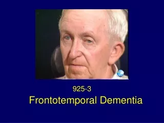
Frontotemporal Dementia (FTD). Monica K. Crane, MD Associate Director Cole Neuroscience Center, UTMCK Clinical Assistant Professor, Dept. of Medicine. Frontotemporal dementia (FTD) Overview. Background and clinical definition Prevalence Anatomy FTD clinical subtypes

Frontotemporal Dementia (FTD)
E N D
Presentation Transcript
Frontotemporal Dementia (FTD) Monica K. Crane, MD Associate Director Cole Neuroscience Center, UTMCK Clinical Assistant Professor, Dept. of Medicine
Frontotemporal dementia (FTD) Overview • Background and clinical definition • Prevalence • Anatomy • FTD clinical subtypes • Neuropathology and genetics of Frontotemporal lobe dementia (FTLD) • Historical cases
FTD = a clinical neurodegenerative disease affecting frontal & temporal lobes • Personality changes • Loss of socially acceptable behavior & emotions • Bizarre and compulsive behaviors • Language dysfunction • Movement disorder
FTD International Research Criteria: Three of the following: Either #7 or #8 one symptom from #1-6 7. Frontal and/or anterior temporal atrophy; other radiologic findings 8. Presence of a known mutation OR • Early disinhibition • Early apathy, loss of motivation • Loss of emotional recognition • Perseverative, compulsive, ritualistic behavior • Hyperorality/ dietary changes • FTD neuropsychological profile B. L. Miller, C. Ikonte, M. Ponton, et al. Neurology 1997;48;937
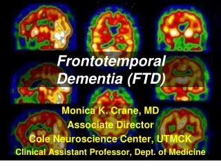
“Dementia That's Neither Alzheimer's Nor Easy” Normal Alzheimer's FTD FDG-PET images of metabolic activity: healthy controls, AD, and FTD. Scale red (high FDG uptake)-yellow-green-blue (low FDG uptake). Photo Credit: Dr. Janet Miller, Dr. Suzanna Lee, MGH/ Harvard, Radiology Rounds April 2006
Dementia Prevalence(% of each type seen in US) FTD syndromes~10-15% Alzheimer’s Disease (AD) ~ 50-70% Vascular dementia~ 5-10% Dementia with Lewy Bodies & Parkinson’s disease dementia~10% • Boxer AL, Miller BL. Alzheimer Dis Assoc Disord. 2005;19 S1:S3-6
FTD Prevalence FTD: Alzheimer’s disease (AD) ratio is 1:1 in those aged 45-65. Ratnavalli et al, Neurology 2002. FTD is more common that AD below age 60. Knopman et al, Neurology 2004. FTD spectrum comprises near 15% or more of the total FTD dementia cases. Boxer AL, Miller BL. Alzheimer Dis AssocDisord. 2005.
Pick’s disease ≠ FTD Pick’s is an autopsy finding only so do not use this term. The clinical disease is FTD. In 1892, Dr. Pick reported a case of a 71 year-old man with focal atrophy and aphasia, & concluded that “progressive brain atrophy can lead to symptoms of local disturbance through local accentuation of the diffuse process.” Dr. Arnold Pick (1851-1924) Prof. of Psychiatry, Prague History of Psychiatry v. 1994, 539-547. GE Berrios , DM Girling. Classic Text No 20. Cambridge.
Frontotemporal lobar degeneration (FTLD) = Neuropathology of clinical FTD Pick’s is a small subset of FTLD From: LM Shaw LM, Korecka M, Clark CM, Lee VMY, Troganowski. Biomarkers of neurodeneration for diagnosis and monitoring therapeutics Nature Reviews Drug Discovery. 2007;6:295-303.
Heterogeneity of FTD subtypes: Anatomy and Clinical presentation
FTD damages 3 major networks:Dorosolateral prefrontal cortex (DLPFC)Anterior cingulate cortex (ACC)Orbitofrontal cortex (OFC)
Areas affected in FTD versus AD Hagmann P, Cammoun L, Gigandet X, Meuli R, Honey CJ, et al. Entorhinal cortex 2009
Frontotemporal dementia subtypes • Behavior variant (bvFTD) • Semantic dementia (SD) • Progressive nonfluent aphasia (PNFA) • Progressive Supranuclear Palsy (PSP) • Corticobasal degeneration (CBD) • FTD with motor neuron disease (FTD-MND) • ALS/CTE (Chronic Traumatic Encephalopathy) • Boxer AL, Miller BL. Clinical features of frontotemporal dementia. Alzheimer Dis Assoc Disord. 2005;19 S1:S3-6
Behavioral variant (bvFTD) Approximately 60% of patients with any form of FTD have bvFTD. Figure 1. Coronal pathology section showing asymmetric right-sided atrophy (R temporal cortices with widening of the inferior horn of the lateral ventral). R L
Gradual onset Impaired judgment and planning Apathy Impaired insight (anosognosia) Loss of empathy and emotion recognition (alexithymia) Disinhibition Abnormal eating behavior Stereotypical or ritualistic behavior Personal neglect Clinical Features of bv-FTD
Profanity use during letter fluency tasks can differentiate FTD from AD. Ringman JM et al. Cogn Behav Neurol 2010;23:159-64
Muangpaisan W. Geriat Aging. 2007; McKhann MG et al. Arch Neurol 2001; Muangpaisan W et al. Neuro J Thai 2003
>50% of FTD subtypes misdiagnosed as primary psychiatric disease Woolley et al. J Clin Psychiatry. 201; 72(2): 126–133. Figure. % of patients initially misdiagnosed prior to ND diagnosis
Computer Self-Test (CST) as a diagnostic tool • Dougherty JD et al. The computerized self test (CST): an interactive, internet accessible cognitive screening test for dementia. J Alzheimer's Dis 2010 Apr;20:185-95. • Crane MK et al. Distinguishing Frontotemporal dementia from Alzheimer’s disease: A pilot study employing the Computer Self-Test (CST). Dementia GeriatrCognDisord 2010;30:62.
CST Cognitive pattern differentiates AD from FTD Crane, MK et al.Neurology. 2011 Suppl(March) 76;
Frontotemporal dementia subtypes • Behavior variant (bvFTD) • Semantic dementia (SD) • Progressive nonfluent aphasia (PNFA) • Progressive Supranuclear Palsy (PSP) • Corticobasal degeneration (CBD) • FTD with motor neuron disease (FTD-MND) • ALS/CTE (Chronic Traumatic Encephalopathy) • Boxer AL, Miller BL. Alzheimer Dis Assoc Disord. 2005;19 S1:S3-6
LEFT predominance •Language features: fluent speech but loss of semantics (word choice) •Reading declines, numbers intact RIGHT predominance • Severe deficits in understanding emotions; loss of empathy • Difficulty recognizing faces and facial expression Eventually R-sided disease progresses to L with language features, and visa versa SD patients develop bvFTD behaviors Semantic dementia (SD) or temporal variant
VIDEO example of bvFTD with phonetic fluency deficits
Frontotemporal dementia subtypes • Behavior variant (bvFTD) • Semantic dementia (SD) • Progressive nonfluent aphasia (PNFA) • Progressive Supranuclear Palsy (PSP) • Corticobasal degeneration (CBD) • FTD with motor neuron disease (FTD-MND) • ALS/CTE (Chronic Traumatic Encephalopathy) • Boxer AL, Miller BL. Alzheimer Dis Assoc Disord. 2005;19 S1:S3-6
Progressive nonfluent aphasia (PNFA) • 20% of FTD cases • Hesitant, effortful speech; stutter or return of childhood stutter • Anomia, agrammatism, sound errors (“gat” for “cat”) • Eventually develop severe movement disorder that overlaps with PSP and CBD Marcel Ravel, (1875-1937) French composer. - in the early stages of PNFA/FTD when composing the orchestral work Boléro (1928).
Fig. Coronal T1 weighted MRIof mild and moderate PNFA Case 1: mild PNFA, atrophy of temporal lobe & pars triangularis. Case 2: moderate PNFA, global atrophy with L-sided and perisylvian predominance. Progressive nonfluent aphasia (PNFA) Case 1 Case 2
Frontotemporal dementia subtypes • Behavior variant (bvFTD) • Semantic dementia (SD) • Progressive nonfluent aphasia (PNFA) • Progressive Supranuclear Palsy (PSP) • Corticobasal degeneration (CBD) • FTD with motor neuron disease (FTD-MND) • ALS/CTE (Chronic Traumatic Encephalopathy) • Boxer AL, Miller BL. Alzheimer Dis Assoc Disord. 2005;19 S1:S3-6
Progressive supranuclear palsy (PSP) • Progressive supranuclear palsy Deterioration of cells in the brainstem, frontal cortex and basal ganglia Dudley Moore 1935-2002
Progressive supranuclear palsy (PSP) key features Postural instability and falls within first year of diagnosis Vertical supranuclear opthalmoparesis Upward gaze paresis with abnormal saccadic eye movements Axial rigidity Cognitive decline Early stage difficult to distinguish from multiple system atrophy, Parkinson disease, and small vessel diease. Most patients with PNFA have PSP or CBD postmortem
PSP radiologic features Hypometabolism on FDG-PET in basal ganglia, brainstem, and frontal lobes
Midbrain atrophy in PSP • (A) Normal: convex upper border of the midbrain • (B) Severe atrophy of the midbrain with • (C) concave upper border of midbrain “humming bird sign”. B A C
Frontotemporal dementia subtypes • Behavior variant (bvFTD) • Semantic dementia (SD) • Progressive nonfluent aphasia (PNFA) • Progressive Supranuclear Palsy (PSP) • Corticobasal degeneration (CBD) • FTD with motor neuron disease (FTD-MND) • ALS/CTE (Chronic Traumatic Encephalopathy) • Boxer AL, Miller BL. Alzheimer Dis Assoc Disord. 2005;19 S1:S3-6
Corticobasal Degeneration (CBD) criteria Core Features Supportive Features Lateralized cognitive dysfunction with preserved memory and learning MRI with asymmetric atrophy in parietal and frontal cortex FDG-PET decreased glucose uptake in parietal and frontal cortex, basal ganglia and thalamus. • Cortical dysfunction • Asymmetric ideomotor apraxia • Alien limb phenomenon • Visual or sensory hemineglect • Focal or asymmetric myoclonus • Non-fluent aphasia (overlap with PNFA) • Extrapyramidal dysfunction • Asymmetric rigidity lacking sustained levodopa response, and focal dystonia
Figure. CBD. Pt1: Mild, focal atrophy of corpus callosum with mild hypometabolism in L frontoparietal cortex (arrow). Pt2: Moderate atrophy of corpus callosum, moderate hypometabolism in L frontoparietal cortex (arrows) Pt3: Severe, diffuse atrophy with bilateral hypometabolism accentuated in the right frontoparietal cortex (arrows)
Frontotemporal dementia subtypes • Behavior variant (bvFTD) • Semantic dementia (SD) • Progressive nonfluent aphasia (PNFA) • Progressive Supranuclear Palsy (PSP) • Corticobasal degeneration (CBD) • FTD with motor neuron disease (FTD-MND) • ALS/CTE (Chronic Traumatic Encephalopathy • Elevated levels of the TDP-43 protein have been found in CTE, a also been identified in patients with CTE, a condition that often mimics ALS and that has been associated with athletes who have experienced multiple concussions and head injury.
FTD with motor neuron disease (FTD-MND) FTD-MND is a CLINICAL PHENOTYPE: • 15% of FTD patients also have FTD-MND • FTD-MND co-occurs in patients with bvFTD but rare in PNFA, CBD, PSP • Early cognitive and behavioral changes with MND symptoms: • slurring of speech, difficulty swallowing, choking • Autonomic dysfunction • limb weakness or muscle wasting • Patients live ≈ 1.4 years after diagnosis (respiratory complications of bulbar symptoms as cause of death) • Most common MND is amyotrophic lateral sclerosis (ALS); older ALS patients may also have behavioral or cognitive problems similar to those seen in FTD (FTD-ALS syndrome)
Results of MRI voxel-based morphometry analyses: behavior & language dominant FTD-MND analysis compared to control Both with frontotemporal gray matter loss. Behavioral variant FTD-MND↓ ↓ in frontal lobes. Language variant FTD-MND ↓ ↓ in L lateral inferior temporal lobe and R putamen. Coon E et al. Neurology 2011;76:1886-1892
Neuropathology and Genetics
FTD inheritance Genetic (40%) Sporadic (60%) 50-80% of individuals appear to be the first person with FTD in the family, also called sporadic or nonfamilial FTD (family not at risk). • Approximately 20-50% of FTD patients have an affected 1st degree relative. • Familial FTD is suspected when 2+ family members are affected in 2+ generations. • Among individuals with FTD, approximately 10% have a single gene mutation (autosomal dominant inheritance).
Frontotemporal Lobar Degeneration (FTLD) is the pathologic confirmation of clinical FTD FTLDs are histopathologic diagnosis with neuronal loss & gliosis, spongiosis & ballooned neurons (image below). Abnormal protein inclusions in neurons & glial cells. • Tauopathies: FTLD with tau+ inclusions • TDP-43 proteinopathies: FTLD with tau-, alpha-synuclein- inclusions which contain the protein TDP-43 + conjugated with ubiquitin+ • FUS: tau-, ubiquitin+, TDP-43-, with fused in sarcoma (FUS) inclusions
Seelar H, Rohrer LD, Pijnenburg YAL, Fox NC, can Swieten JC. Clinical, genetic and pathological heterogeneity of frontotemporal dementia: A review. J Neurol Neurosurg Psychiatry 2010.