Download
1 / 45
450 likes | 584 Vues
Explore the role of dopamine in motivation, reward, and addiction, as well as the enzymes involved in its breakdown. Learn about neurotransmitter metabolism and the impact of drugs on neurotransmitter release.

E N D
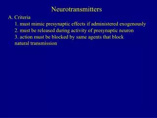
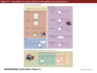
6.1 Examples of small-molecule and peptide neurotransmitters. (Part 1)
6.1 Examples of small-molecule and peptide neurotransmitters. (Part 2)
6.1 Examples of small-molecule and peptide neurotransmitters. (Part 3)
6.1 Examples of small-molecule and peptide neurotransmitters. (Part 4)
6.1 Examples of small-molecule and peptide neurotransmitters. (Part 5)
6.15 Neuropeptides vary in length, but usually contain between 3 and 36 amino acids.
5.5 Metabolism of small-molecule and peptide transmitters. (Part 1)
5.5 Metabolism of small-molecule and peptide transmitters. (Part 3)
5.5 Metabolism of small-molecule and peptide transmitters. (Part 4)
5.12 Differential release of neuropeptide and small-molecule co-transmitters.
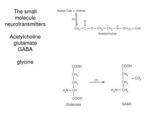
6.2 Acetylcholine metabolism in cholinergic nerve terminals.
6.6 Glutamate synthesis and cycling between neurons and glia.
6.8 Synthesis, release, and reuptake of the neurotransmitters GABA and glycine. (Part 2)
6.8 Synthesis, release, and reuptake of the neurotransmitters GABA and glycine. (Part 1)
Figure 6.10 The biosynthetic pathway for the catecholamine neurotransmitters
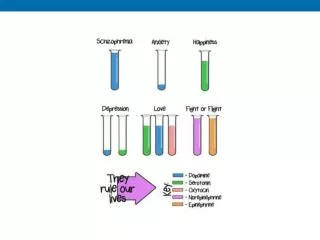
Neurotransmitters and transporters: Dopamine factoids • Dopamine is believed to be involved in motivation, reward, and reinforcement and many drugs of abuse affect dopaminergic synapses. Dopamine action in the synaptic cleft is terminated by the reuptake of dopamine into the nerve terminal or neighboring glia by a Na+ dependent dopamine transporter (DAT). Cocaine exerts its effects by binding to and inhibiting DAT which causes an increase in the amount of dopamine in the synaptic cleft. • Amphetamine, another addictive drug, also inhibits DAT as well as the transporter for norepinephrine. • The two major enzymes involved in the breakdown of dopamine are monoamine oxidase (MAO) and catechol O-methyltransferase (COMT). Inhibitors of these enzymes such as phenelzine and tranylcypromine are used clinically as antidepressants.
5.22 A neurotransmitter can affect a postsynaptic cell via two types of receptor proteins. (Part 2)
Figure 7.6 Effector pathways associated with G-protein-coupled receptors
5.22 A neurotransmitter can affect a postsynaptic cell via two types of receptor proteins. (Part 1)
6.4 The general architecture of ligand-gated receptors. (Part 1)
The families of ligand-gated receptors. Glutamate receptor subtypes are named after the agonists that activate them: AMPA: a-amino-3-hydroxyl-5-methyl-4-isoxazole-propionate NMDA: N-methyl-D-aspartate Kainate: Kainic acid Most central synapses have both AMPA and NMDA receptors
GABA acting on GABAARs usually hyperpolarizes cells because ECl is more negative than the resting potential, so Cl- flows into the cell. In some cases GABA can evoke depolarization instead of hyperpolarization in neurons. This can occur if the chloride equilibrium potential (ECl) is positive to the resting potential. What controls where ECl is? It depends on intracellular chloride concentration, which appears to be regulated primarily by two transporters, an Na-K-Cl cotransporter (NKCC) that normally accumulates chloride and a K-Cl cotransporter (KCC) that normally extrudes chloride (for review, see Russell, 2000). Certain neurons and epithelial cells with ECl positive to the resting potential express NKCC, whereas neurons with an ECl negative to the resting potential express KCC2 (the neuron-specific isoform).
Figure 6.6 Postsynaptic responses mediated by ionotropic glutamate receptors (Part 2)
Figure 5.17 Activation of ACh receptors at neuromuscular synapses
Figure 5.18 The influence of the postsynaptic membrane potential on end plate currents EPC=g(Vm-Erev)
5.16 The influence of the postsynaptic membrane potential on end plate currents. (Part 3) Lower external Na+ Higher external K+
5.17 The effect of ion channel selectivity on the reversal potential. (Part 1) EPC=g(Vmem – Erev)
5.17 The effect of ion channel selectivity on the reversal potential. (Part 2)
5.19 Reversal and threshold potentials determine postsynaptic excitation and inhibition. (Part 1)
5.19 Reversal and threshold potentials determine postsynaptic excitation and inhibition. (Part 2)