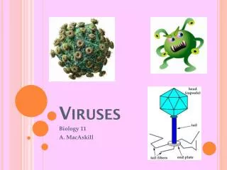
Viruses
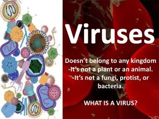
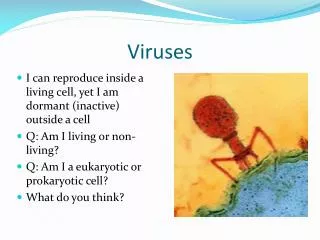
Viruses. General Characteristics of viruses. 1. Depending on one’s viewpoint, viruses may be regarded as exceptionally complex aggregations of nonliving chemicals or as exceptionally simple living microbes. How does it differ from a cell?

Viruses
E N D
Presentation Transcript
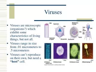
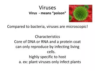
General Characteristics of viruses 1. Depending on one’s viewpoint, viruses may be regarded as exceptionally complex aggregations of nonliving chemicals or as exceptionally simple living microbes. How does it differ from a cell? 2. Viruses contain a single type of nucleic acid (DNA or RNA) and a protein coat, - sometimes enclosed by an envelope composed of lipids, proteins and carbohydrates. They are not cells or composed of cells.
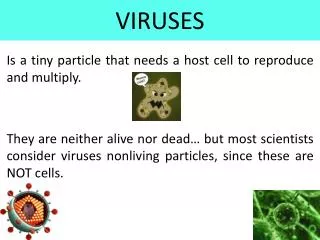
General Characteristics of viruses 3. Viruses are obligatory intracellular parasites. 4. A virion is a complete, fully developed viral particle composed of nucleic acid surrounded by a coat.
Host Range • Host range refer to the spectrum of host cells in which a virus can multiply. 2. Most viruses infect only specific types of cells in one host species. 3. Host range is determined by the specific attachment site on the host cell’s surface and the availability of host cellular factors.
Viral Size 1. smaller than bacteria. 2. Viruses rage from 20 to 14,000 nm in length.
Classification of Viruses 1. Classification of viruses is based on type of nucleic acid, morphological class, and presence or absence of an envelope. 2. Virus family names end in -viridae; genus names end in -virus; specific epithets have not been assigned. 3. A viral species is a group of viruses sharing the same genetic information and ecological niche.
Nucleic Acid 1. Viruses contain either DNA or RNA, never both, and the nucleic acid may be single- or double- stranded, linear or circular, or divided into several separate molecules. DNA or RNA SS or DS Linear or circular or divided
Capsid • The protein coat surrounding the nucleic acid of a virus is called the capsid. 2. The capsid is composed of subunits, capsomeres, which can be a single type of protein or several types.
General Morphology 1. Helical viruses (for example, tobacco mosaic virus) resembling long rods, and their capsids are hollow cylinders surrounding the nucleic acid.
Polyhedral viruses 2. Polyhedral viruses (for example, adenovirus) are many-sided. Usually the capsid is an icosahedron. 20 triangular faces 12 corners example polio virus
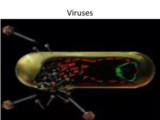
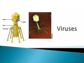
Complex viruses 3. Complex viruses have complex structures. For example, many bacteriophages have a polyhedral capsid with a helical tail attached.
Capsid and Envelopes 3. The capsid of some viruses is enclosed by an envelope consisting of lipids, proteins, and carbohydrates. 4. Some envelopes are covered with carbohydrate-protein complexes called spikes. Attachment, absorption. Example: influenza
Cultivation of Viruses • Viruses must be grown in living cells (cell culture). Animals and plant host are expensive and not easy to maintain. 2. The easiest viruses to grow are bateriophages (Phage = eater of bacteria). Easy to manipulate bacterial cells and their viruses in the laboratory
Growing Viruses 1. The plaque method mixes bacteriophages with host bacteria and nutrient agar. 2. After several viral multiplication cycles, the bacteria in the area surrounding the original virus are destroyed; the area of lysis is called plaque. 3. Each plaque originates with a single viral particle; the concentration of viruses is given as plaque-forming units (PFU)
Growth of Animal Viruses in the Laboratory • Cultivation of some animal viruses requires whole animals Humans are the test subjects but processes are slow to see the results Simian AIDS (1986) and feline AIDS provide models for study of human AIDS. Genetically engineered mice. SCID –Human mouse 2. Some animal viruses can be cultivated in embryonated eggs
Growth of Animal Viruses in the Laboratory • Cell cultures are cells growing in culture media in the laboratory • Primary cell lines (few generations) cell lines grow for a short time in vitro. • Embryonic cell lines (100 generations). • Continuous cell lines can be maintained in vitro indefinitely. • Transformed or cancerous cells a. HELA 1951
Viral multiplication Bacteriophages
Multiplication of Bacteriophages 1. During a lytic cycle, a phage causes the lysis and death of a host cell. 2. Lysogeny. DNA incorporated as a prophage into the DNA of the host cell
Lytic Cycle • The multiplication cycle of these phages can be divided into five distinct stages: • Attachment • Penetration • Biosynthesis • Maturation • Release
Lytic Cycle: Attachment • During the attachment phase of the lytic cycle, • Chance collision • Sites on the phage’s tail fibers attach to complementary receptor sites on the • bacterial cell.
Lytic Cycle:Penetration • Phage lysozymes opens a portion of the bacterial cell wall, • tail sheath contracts to force the tail core through the cell wall, • DNA enters the bacterial cell and the capsid remains outside.
Lytic Cycle: Biosynthesis, • Phage DNA is replicated • Phage DNA produces mRNA coding for proteins necessary for phage multiplication • capsids and proteins are produced
Lytic Cycle: Maturation • Phage DNA and capsids are assembling into complete viruses
Lytic Cycle: Release (lysis) • phage lysozyme breaks down the bacterial cell wall, and the multiplied phages are released
Vocabulary • Burst time: The time from phage attachment to release (AVG 20 to 40 min). • Burst size: The number of newly synthesized phages from a single infected cell (50-200). • Eclipse period The time period when whole virons can not be found. It is the time from the end of penetration to the beginning of release.
Lysogeny • Some viruses (lysogenic phages) do not always cause lysis and death of the host cell when they multiply. • These viruses may incorporate their DNA into the host cell’s DNA to begin a lyogenic cycle. • In lysogeny, the phage remains latent or inactive
Characteristics of lysogeny • Lysogenic cells are immune to reinfection by the same phage. • Repressor proteins stop transcription of all other phage genes. • Host cell may exhibit new properties (phage conversion) • Bacteria may acquire new genes from previously infected cells • Special transduction
Generalized Transduction • Information is transported from one bacteria to another via a phage. • Bits of host DNA are packaged along with the phage DNA in the capsid head. • Unlike specialized transduction the transported gene does not have to lie adjacent to the prophage on the host chromosome. • The host gene is randomly picked up in the cytoplasm after the chromosome has been degraded.
Animal Viruses • The multiplication cycle of these phages can be divided into six distinct stages: • Attachment • Penetration • Uncoating • Biosynthesis • Maturation • Release
Multiplication of a DNA Papovavirus Viral DNA enters Cell’s nucleus Enzymes synthesized for DNA replication
RNA Picornaviruses sense strand (+strand) virus
Things to know for the Exam Animal virus life cycle