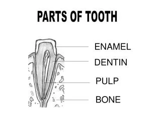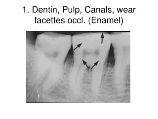
Dentin, Enamel, Pulp
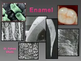
Dentin, Enamel, Pulp. Enamel. most highly mineralized tissue 96% mineral, 4% water inorganic crystalline calcium phosphate – hydroxyapatite various ions like strontium, magnesium, lead and fluoride are present at some point during enamel formation

Dentin, Enamel, Pulp
E N D
Presentation Transcript
Enamel • most highly mineralized tissue • 96% mineral, 4% water • inorganic crystalline calcium phosphate – hydroxyapatite • various ions like strontium, magnesium, lead and fluoride are present at some point during enamel formation • enamel is very brittle but the underlying dentin provides some resilience
Enamel • organized into rods (prisms) and interrods (interprismatic substance) • built from closely packed ribbon-like hydroxyapatite crystals – 60 to 70 nm in width and 25 to 30 nm in thickness • the calcium phosphate unit has hexagonal symmetry that are stacked together as rod or interrod enamel • the rod is shaped somewhat like a cylinder and is made up of crystals that organize with their long axes parallel to the longitudinal axis of the entire rod • this organization is tighter around the center of each rod • the interrod region surrounds each rod – its crystals are oriented along different axes from the rod
Enamel • the boundary between rod and interrod is delimited by a narrow space containing organic material – rod sheath
Amelogenesis • two-step process • first step produces a partially mineralized enamel – approximately 30% mineralized • second step involves an influx of additional mineral content – coincident with the removal of organic material and water – results in 96% mineralization • the influx of mineral results in growth of the crystals in width and thickness
Amelogenesis • ameloblasts – derived from the inner dental epithelium • secrete matrix proteins that are responsible for creating and maintaining and extracellular environment favorable to mineral deposition • possess a unique life cycle – each stage reflects its primary activity during enamel stages • can be divided into three main stages • presecretory, secretory, maturation stages
Amelogenesis Figure 7-14 Representative micrographs of amelogenesis in the cat. A, Tooth formation shows an occlusal-to-cervical developmental gradient so that on some crowns finding most of the stages of the ameloblast life cycle is possible. The panels on the right (B corresponds with B1 and C with B2) are enlargements of the boxed areas: B, Secretory stage, initial enamel formation; C, secretory stage, inner enamel formation. D and E are from the incisal tip of the tooth (see Fig. 7-15). D, Midmaturation stage, smooth-ended ameloblasts; and E, late maturation stage, ruffle-ended ameloblasts. Am, Ameloblasts; D, dentin; E, enamel; N, nucleus; Od, odontoblasts; PL, papillary layer; RB, ruffled border; SB, smooth border; SI, stratum intermedium.
Presecretory stage • morphogenetic phase • during the cell stage – shape of the crown is determined • separated from the dental papilla by a basement membrane • differentiation phase • as the cells of the IDE differentiate into pre-ameloblasts they elongate and their nuclei shift toward the stratum intermedium • the basement membrane fragments by the cytoplasmic projections of the pre-ameloblasts – during the formation of predentin • this allows contact between the IDE/pre-ameloblasts and dental papilla • at the distal end of the cell – extensions form called Tome’s processes (proximal portion) - against which enamel forms • adjacent ABs align closely with each other – form junctional complexes between them keeps them aligned
Secretory Stage • Pre-AB cells acquire intense synthetic and secretory activity • enamel proteins are translated by the RER, modified by the Golgi and packaged into secretory granules • secretion is constitutive – the secretory granules are not stored for long within the cells • the contents of the secretory granules are released against the newly formed dentin along the surface of the Tome’s process • little time elapses between the secretion of enamel and its mineralization • as the initial enamel layer forms – the ABs migrate away from the dentin surface and develop a distal portion of Tome’s process – extension from the existing proximal portion of Tome’s process • the pre-AB only has a pTP during initial enamel formation • the dTP interdigitates into the enamel beyond the pTP • the dTP lengthens as the enamel layer thickens • it also becomes thinner as the rod grows in diameter • eventually squeezed out of existence – enamel in this area is known as a rod
Figure 7-31 In three dimensions, interrod (IR) enamel surrounds the forming rod (R) and the distal portion of Tomes’ process (dpTP); this portion is the continuation of the proximal portion (ppTP) into the enamel layer. The interrod (IGS) and rod (RGS) growth sites are associated with membrane infoldings (im) on the proximal and distal portions of Tomes’ process, respectively. These infoldings represent the sites where secretory granules (sg) release enamel proteins extracellularly for growth in length of enamel crystals and, consequently, the thickening of interrod and rod enamel.
Maturation Stage • Maturation of enamel and maturation of pre-AB into mature ABs • before tooth eruption – the enamel hardens • the pre-existing HA crystals of the enamel grow in width and thickness and NOT because new crystals are made • Tome’s processes are not apparent at this stage • the ABs are generally referred to as post-secretory cells at this stage • although they still secrete proteins • made up of a transitional phase and the maturation proper phase • transitional phase – after the full thickness of the enamel has formed • reduction of AB height and a decrease in their volume and organelle content • maturation proper phase – ABs become involved in the removal of water and organic material • also undergo apoptosis so that approximately 25% of the ABs die during the transitional phase and an additional 25% die during the MP phase • characterized by the modulation of the cells – cyclic creation, loss and recreation of a highly ruffled apical surface and a smooth surface • significance is unknown – could be related to calcium transport and completion of enamel mineralization • organic matrix removal is the production of bulk-degrading enzymes into fragments small enough to be able to leave the enamel layer and be taken up by the AB
Enamel proteins • amelogenin – accumulate during the secretory stage • undergo minor short-term and major long-term processing to form smaller fragments • these fragments form the bulk of the final organic matrix of maturing enamel • prevents crystals from fusing during their formation and must be removed to permit crystal growth • nonamelogenins • enamalin – small degredation during the secretory stage which decreases in the deeper zones of the enamel • crystal nucleation and growth • ameloblastin – undergoes rapid degredation – the intact protein is found near the enamel-forming surface while the fragmented forms are found in the deeper zones of the enamel • promotes mineral formation and crystal elongation • tuftelin – localizes specifically at the DEJ and participates in its establishment • not specific to enamel • enzymes: metalloproteinases – e.g. MMP20 or enamelysin (short-term breakdown), serine proteinases (bulk degredation), phosphatases • dentin sialoprotein – transiently expressed
Mineral pathway • introduction of minerals span the secretory and maturation phases • calcium moves from the blood supply through the enamel organ to reach the enamel • extracellular source • a smooth tubular network has been described that is found in ABs and opens onto the enamel • this network is similar to the ER/sarcoplasmic reticulum • calcium is likely to be routed from high-capacity stores associated with the ER • intracellular source • almost an immediate formation of crystallites within the enamel secreted against the dentin – so there is no pre-enamel
Enamel structural organization • Striae of Retzius – in longitudinal sections they are a series of dark lines extending from the DEJ toward the tooth surface • Cross striations – forms at 4um intervals across the rods • rhythmicity?? • Bands of Hunter and Schreger – optical phenomenon produced by the changing orientations of adjacent groups of rod • Gnarled enamel • Enamel tufts and lamellae – no clinical significance • like geological faults • project from the DEJ for a short distance into the enamel • branched and contain greater concentrations of enamel that the rest of the enamel • abrupt changes in the directions of the rods arising from the DEJ
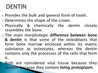
Dentin – basic structure • first deposited as a layer of pre-dentin – unmineralized matrix • principally collagen and noncollagenous components – similar to osteoid • has an elastic quality that is important to the functioning of the tooth – the elasticity provides flexibility and prevents fracture of the overlying more brittle enamel • has a yellowish color • gradually mineralizes to form dentin – as noncollagenous proteins are incorporated at the mineralization front • mature dentin – 70% inorganic, 20% organic, 10% water by weight • inorganic – hydroxyapatite in the form of small plates • organic – collagen types I, III and V with small amounts of lipids and noncollagenous matrix proteins • the NCMPs pack the space between the CN fibers and accumulate along the periphery of the dentinal tubules • dentin phosphoprotein, sialoprotein, matrix protein, osteonectin/SPARC, osteocalcin, osteopontin, proteoglycans and some serum proteins • CN type I acts as a scaffold for the accomodation of a large amount of mineral in the holes and pores of the fibrils • the NCMPs regulate mineral deposition along CNI fibrils – acting as activators or inhibitors
Types of Dentin • Primary dentin • forms most of the tooth • when it outlines the pulp chamber it is referred to as circumferential dentin • outer layer – mantle dentin (mineralized differently) • Secondary dentin • develops after root formation has been completed • has a tubular structure that is continuous with the primary dentin • dentinal tubules are less organized the primary dentin – some regions may lack tubules or some may regions may have thicker tubules • ratio of mineral to organic is the same as in primary dentin • not deposited evenly along the periphery of the pulp chamber (especially in the molars) • greater deposition along the roof and floor of the chamber leads to an asymmetric reduction in the chambers size and shape = pulp recession • Tertiary dentin • reactive or reparative dentin • produced in reaction to various stimuli – attrition, restorative procedures • produced only by those cells affected by the stimulus • may have tubules – sparse in numbers and irregularly arranged • deposition is very rapid – ODs can become trapped in the dentin and distorts the tubular pattern • little is known of the deposition process – collagen fibers= production appears to be downregulated
Pattern of dentin formation • begins at the bell stage • in the papillary tissue adjacent to the folded IDE – spreads down the cusp slope as far as the cervical loop of the enamel/dental organ • root dentin forms slightly later – require the formation of Hertwig’s rooth sheath from the CL • rates of dentin deposition vary with tooth but also can very within each tooth • continues throughout the life of the tooth – progressive reduction of the pulp chamber
Dentinogenesis • Odontoblast differentiation • differentiate from the mesenchyme of the dental papilla • occurs through the interaction with the IDE • mirror image in polarity when compared to the differentiating pre-ABs • Formation of Mantle dentin • after OD differentiation – the dental matrix forms • ODs differentiate in the pre-existing ground substance of the dental papilla • the first collagens are deposited in the DP • these first CN fibers are large-diameter = Korff’s fibers (CNIII + fibronectin) • as the ODs increase in size and mature - produce smaller CNI fibrils that orient themselves parallel to the future DEJ • also develops a process = Tome’s fiber – left behind in the forming dentin matrix as the OD moves away from the BM toward the forming pulp • Vascular supply • when the mantle dentin forms, capillaries are found beneath the newly differentiated OD layer • as circumpulpal dentin forms – these capillaries migrate between the ODs outer cell OD inner cell cells of DP inner cell Figure 8-7 Odontoblast differentiation. The undifferentiated cells (A) of the dental papilla divides (B), with its mitotic spindle perpendicular to the basement membrane. A daughter cell (C), influenced by the epithelial cell (D), differentiates into an odontoblast (E).
Control of Mineralization • the mineral phase first appears within the matrix vesicles next to the BM as single crystals • crystals grow rapidly and rupture from the confines of the vesicles and spread as clusters of crystallites that fuse with adjacent ones • deposition lags behind that formation or the organic matrix so that the organic pre-dentin is always found between the ODs and the mineralization front and the crystals spread and fuse • does the OD control this process?? • control is exerted probably through the secretion of non-collagenous matrix proteins
Formation of Root Dentin • compositionally different that coronal dentin • the collagen fibers in the mantle dentin are arranged in a different orientation • the phosphoryn protein content is less • the degree of root dentin is also less • rate of deposition of dentin is slower
Linear Globular Histology • Dentinal tubules • for the passage of OD processes • similar to canaliculi • their configuration is reflective of the course taken by the ODs during dentinogenesis – S-shaped path • Peritubular dentin • tubules are delimited by a collar of more highly calcified matrix = peritubular dentin • about 40% more mineralized than dentin • contains little CN and may be enriced in the non-CN matrix proteins • Intertubular dentin • located between the dentinal tubules • represents the primary secretory product of the ODs • consists of tightly interwoven CNI fibers + apatite crystals • Interglobular dentin • areas of unmineralized or hypomineralized dentin • where globular areas of mineralization have failed Figure 8-21 Scanning electron microscope preparations of predentin (A and B) and dentin (C and D). A and B, Although no dentinal tubules (dt) occur in predentin, each odontoblast process (Odp) is surrounded by intertwined collagen fibrils (Coll) that outline the future dentinal tubule. As visible in cross-sectional (A) and longitudinal (B) profile, the fibrils run circumferentially and perpendicular to the process. C, In healthy dentin each tubule is occupied by a process or its ramifications. D, The dentinal tubule is delimited by a layer of peritubular dentin (arrowheads) that is poor in collagen and more mineralized than the rest of the dentin. The dentin between tubules is referred to as intertubular dentin (iD).
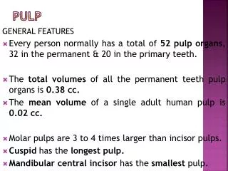
Pulp • soft connective tissue that supports the dentin • four distinct zones • 1. odontoblastic zone at the periphery • 2. cell-free zone of Weil – below the ODs • 3. cell-rich zone • 4. pulp core – major vessels and nerves
Pulp • Odontoblasts • line the periphery of the pulp chamber • columnar in the crown region of the fully developed tooth • more cuboidal at the midpoint of the pulp chamber • the morphology reflects their activity – the more active the more elongated they are – with more cytoplasm • active ODs within the pulp have prominent organelles with multiple vesicles • CN pathway is similar to that of the pulp fibroblasts • CNs and non-CN proteins are packaged into secretory granules for exocytosis • the non-CN proteins are the same as those found in the dentin
Pulp • Fibroblasts • greatest number • numerous in the coronal portion of the pulp • form the cell-rich zone • form and maintain the pulp matrix – CN fibers and ground substance • in young pulp the fibroblasts are active and have extensive cytoplasm and organelles • decrease in size with age and they flatten • Ectomesenchymal cells • undifferentiated cells of the pulp • from neural crest (ectodermal) • depending on the stimulus – give rise to the ODs or fibroblasts of the pulp • Macrophages • Lymphocytes • Dendritic cells
Pulp vascular & nerve supply • enter and exit via the apical foramen as a bundle of vessels and nerves • smaller vessels and nerves enter through the minor foramina • arterioles occupy a central position within the pulp and give off small lateral branches that extend toward the subodontoblastic zone • main capillary beds are located below the ODs • nerves follow the same course as the afferent arterioles • contribute to a plexus in the cell-free zone just below the ODs in the coronal section of the pulp chamber • subodontoblastic plexus of Raschkow • no corresponding plexus can be found in the root – ascending trunks give off branches
Pulp – nervous supply Figure 8-59 Nerve fibril arising from the plexus of Raschkow is shown passing between the odontoblasts and looping within the predentin. (From Bernick S: Innervation of the teeth. In Finn SB, editor: Biology of the dental pulp organ, Tuscaloosa, 1968, University of Alabama Press.) Figure 8-60 Nerve at the predentin-dentin (PD, D) junction demonstrated by staining for nerve growth factor receptor in a tangential section. Its extensive ramification is notable. (From Maeda T, Sato O, Iwanaga T, et al: Immunocytochemical localization of nerve growth factor receptor