Download
1 / 44
450 likes | 476 Vues
Join Dr. Michelle Brazas to explore over 200 types of cancer, how mutations lead to cancer, and prevention steps. Learn hands-on methods to compare normal and cancer cells and extract DNA for analysis. Discover potential treatments based on research findings.

E N D
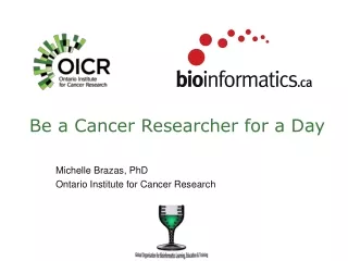
Be a Cancer Researcher for a Day Michelle Brazas, PhD Ontario Institute for Cancer Research
Acknowledgements John McPherson Geoffrey Fong Genome Technologies Team
What is Cancer? Cancers are diseases of unstoppable cell growth Over 200 different types of cancer!
Any normal cell can turn into a cancer cell Esophagus Skin Bladder Kidney Breast Stomach Liver Lung Bone Blood Pancreas Colon Head
How does a normal cell change into a cancer cell? Mutations build up in your DNA DNA mutations change a normal cell into one that grows unstoppably
What is a mutation? Also insertions and deletions of bases. Also translocations (different chromosomes joining together).
What causes mutations in DNA? • Random mutation events in DNA replication • Family Genetics • Life Style and Habits • Environmental You can change your life style You can change your environment Mutations in DNA accumulate over time to cause cancer
What can you do to prevent cancer? • Don’t smoke. • Maintain a healthy weight. • Exercise regularly. • Eat a healthy diet. • Drink alcohol only in moderation, if at all. • 6. Protect yourself from the sun. • 7. Protect yourself from infections. • 8. Get screening tests regularly. From Dart et al, Cancer Causes and Control, 2012
Be a Cancer Researcher Research Problem: Bud has blood cancer. He has come to you for help. He wants a treatment that will get rid of his blood cancer, but won’t make him more sick. To help Bud, your Research Question is: What is the difference between normal blood cells and Bud’s cancer blood cells? Why do we want to look at the difference?
How can you answer this research question? Possible Experiments that Answer Question: • Compare the normal cells & cancer cells under the microscope What is the difference between normal blood cells and Bud’s cancer blood cells?
Making a Blood Smear on a Microscope Slide http://www.youtube.com/watch?v=O3d_4dkVVSE&feature=youtu.be
Staining a Blood Smear http://www.youtube.com/watch?v=89VRmOJ10iA&feature=youtu.be
Microscope Comparison - Pathology Normal Blood Cells Cancer Blood Cells Chronic myeloid leukemia Can we give Bud a treatment based on this answer? What is the difference between normal blood cells and Bud’s cancer blood cells?
How can you answer this research question? Possible Research Methods to Answer Question: • Compare the normal cells & cancer cells under the microscope • Compare the DNA from the normal & cancer cells What is the difference between normal blood cells and Bud’s blood cancer cells?
DNA To compare the DNA between normal & cancer cells we must separate the DNA from the rest of the cell
DNA Extraction from Cheek Cells (20min) Step 1 • Pour 3mL of TheraBreath mouthwash into your cup. Step 2 • Swirl the mouthwash in your mouth for 30 seconds. • Gently bite on your cheeks. • Spit the mouthwash back into your cup. Step 3 • Pour the mouthwash with cheek cells into the 15mL tube. • Discard the cup into the yellow waste bag. Step 4 • Using the bulb pipette, add 1mL of soap solution to the tube. • Gently mix by inversion.
DNA Extraction from Cheek Cells Step 5 • Layer cold isopropanol on top of the soap solution by slowly pouring down the side of the tube to the 15mL mark. Step 6 • Let the solution sit for 5 minutes to separate. • DNA will precipitate into the alcohol layer. Step 7 • Use the stir stick to play with your DNA. DNA dissolves in water but precipitates in alcohol.
To visualize our DNA, we need to run it on an agarose gel http://www.youtube.com/watch?v=2UQIoYhOowM&feature=youtu.be
DNA Comparison – Molecular Biology • Stain the agarose gel to view the DNA • Lane M – Lambda DNA/HindIII Ladder (for sizing) • Lane 1 – Normal Blood DNA • Lane 2 – Cancer Blood DNA What is the difference between normal blood cells and Bud’s cancer blood cells? Can we give Bud a treatment based on this answer?
Can also visualize DNA by Spectral Karotype – Molecular Genetics Normal Blood Cell Karotype Cancer Blood Cell Karotype What is the difference between normal blood cells and Bud’s cancer blood cells?
How can you answer this research question? Possible Research Methods to Answer Question: • Compare the normal cells & cancer cells under the microscope • Compare the DNA from the normal & cancer cells • Sequence the DNA bases & compare the DNA of the normal & cancer cells What is the difference between normal blood cells and Bud’s cancer blood cells?
Sequencing DNA really fast http://www.youtube.com/watch?v=HtuUFUnYB9Y&feature=youtu.be
Results from the sequencer… 160 million short reads of DNA OR Total of 10 billion DNA bases • To understand the information in the DNA sequence reads, we need to assemble them in the proper order • Then we can compare normal DNA sequence to cancer DNA sequence
The Sequence Assembly Race (30min) Work in your Team: • One DNA sequence read will overlap with another DNA sequence read by a few bases. • Assemble all of the sequence reads together. • Tape them together to secure them. (Computers are used to assemble 160 million sequence reads!)
Getting Information from a Sequence Alignment • After assembling the tumor DNA and assembling the normal DNA, the two assemblies are compared • From a sequence alignment of cancer DNA to normal reference DNA, we can see where mutations occur Mutations Sequence reads from Cancer DNA Normal DNA sequence Position: 1 2 3 4 5 6 7 8 9… • 2 sequence reads have the same bases as the normal DNA sequence • + 2 reads have different bases compared to the normal DNA sequence What is the difference between normal blood cells and Bud’s cancer blood cells?
DNA is the instruction manual for making proteins. If DNA is mutated, then usually the protein is also mutated.
What is the impact of Bud’s mutations? Knowing that mutations exist is only useful information if we know what cellular function they change • Do Bud’s mutations occur outside of important sections (protein coding sections) in the DNA? • Mutations here might not change anything in the cell • Do Bud’s mutations occur within an important gene, like a gene that controls cell growth? • Mutations here might turn off the control, allowing the cell to keep growing without stopping
Determining Location of Mutations with BLAST Step 1 • Go to http://goo.gl/6E7XoF Step 2 • Select a sequence file of your choice. This sequence comes from our read assembly activity. • Open the sequence file. • Select (Ctrl+A) and copy (Ctrl+C) all of the sequence. Step 3 • Go to http://blast.ncbi.nlm.nih.gov/ • Select ‘Human’ under BLAST Assembled RefSeq Genomes. Step 4 • Paste (Ctrl+V) the copied sequence into the ‘Query Sequence’box. • Hit to start the comparison (alignment) of your DNA sequence to the whole Human genome sequence. • BLAST will return locations in the Human genome that match (align to) your input sequence.
Determining Location of Mutations with BLAST Step 5 • Which chromosome (or chromosomes) is your sequence is located? • Hover over each of the red lines to determine the chromosome number. Step 6 • In the ‘Descriptions’ box, select the top result. This jumps down the page to the result. • Download the ‘Graphics’for this result. This opens a new ‘Graphics’ tab. • Note: Your sequence may match to more than one chromosome so you will need to repeat this step for each chromosome. Step 7 • A new ‘Graphics’ window opened. • In the ‘Sequence’ section, which Gene does your sequence match with? Step 8 • Hover over the gene: What is the gene title, location, and length? • Select ‘View MIM’from the pop up window to learn about the function of your gene. • Learn about the function of your gene in the ‘Gene Function’ section.
Answers to BLAST • Sequence #1: • Chromosome 22 = BCR gene • Chromosome 9 = Abl1 gene • Sequence #2: • Chromosome 22 = BCR gene • Sequence #3: • Chromosome 9 = Abl1 gene
Impact of Mutations on Bud’s Blood Cells • Normal Abl1 protein (on chromosome 9) is a cell growth factor • In chronic myeloid leukemia (CML), DNA mutates so that chromosome 9 + chromosome 22 exchange DNA segments • A mutation fuses DNA to create BCR (Chr. 22) + Abl (Chr. 9) = BCR/Abl • Mutant BCR/Abl protein never turns off • The result is that the blood cell receives instructions to “Keep Growing!” Mutant fusion: BCR/Abl gene Common mutation in Chronic Myeloid Leukemia (CML)
Making a Drug to Stop the Mutant Protein • Scientists could work with this information to model the normal Abl protein and compare it to the mutant BCR-Abl protein • The goal is to design a drug that works against the BCR-Abl protein • Need to look at the 3D structure of Abl protein • Need to look at how a drug interacts with this protein in 3D
3D Model of Abl and Gleevec (STI-571) Corbin A S et al. J. Biol. Chem. 2002;277:32214-32219 Using a 3D model of the Abl protein with Gleevec, we can determine which amino acids are important for Gleevec to work as a cancer drug
Using 3D Protein Models of Abl + Gleevec Step 1 • Go to http://www.pdb.org/pdb/home/home.do Step 2 • Search for 1IEP • Under the image, click on 3D View Step 3 • Select Custom View • Jmol mode = WebGL (beta) • Style = Backbone (or Ligands and Pocket) Step 4 • Rotate the 3D model • See how Gleevec fits into the pocket of the Abl protein
Using 3D Protein Models of Abl + Gleevec Step 5 • Choose the Abl amino acids that you think are important for interaction with Gleevec (look at the figure for the chemical model of Abl and Gleevec) Step 6 • Under the 3D image, expand the Scripting Options box • Type in the Input box (the numbers are the amino acid numbers you chose to be important): select 271, 286, 290, 315, 381; spacefill; color yellow; Step 7 • To color Gleevec, type in the Input box: select ligand; spacefill; color red; Step 8 • Why is amino acid 310 not important to Gleevec function in Abl? • Type in the Input box: select 310; spacefill; color blue;
11 amino acids (yellow) within tyrosine kinase domain of Abl protein are important for interacting with Gleevec (red)
Can we give Bud a treatment based on this answer? YES http://www.dnalc.org/view/15055-Using-DNA-science-to-control-CML-Brian-Druker.html
How Gleevec works on Mutant BCR/Abl • Goal of cancer drug is to stop the activity of mutant BCR/Abl protein • Proteins have domains. The important domain in mutant BCR/Abl is tyrosine kinase. • Tyrosine kinase domain moves phosphates (from ATP) around • The tyrosine kinase domain takes a phosphate from ATP and transfers it to the tyrosine amino acid on a substrate • The phosphated substrate then passes along the message to keep growing • Mutant BCR/Abl is stuck on: Tyrosine kinase domain is always transferring a phosphate, so there is always a message to keep growing Phosphate ATP – Adenosine Triphosphate
Stopping the Activity of Mutant BCR/Abl • Need a molecule that can block ATP from entering the tyrosine kinase domain of the mutant BCR/Abl protein • No ATP = tyrosine kinase domain cannot transfer phosphates = No more growth message. ATP – Adenosine Triphosphate What is similar about these two molecules?
Gleevec Blocks the Mutant BCR/Abl Activity Always ON Blocked by STI571 (Gleevec) No Phosphate We discovered that the DNA from Bud’s blood cancer is mutated to create the BCR/Abl gene BCR/Abl protein. So we could give Bud this drug to block BCR/Abl activity.
Bud’s Blood Counts Today http://www.dnalc.org/view/15043-Bloodcount-returns-to-normal-with-Gleevec-Bud-and-Yvonne.html
The End Thank you!