
76N-TERT.V
Supplementary Figure 1. 76N-TERT.ADA3. MEF. ADA3 fl/fl. MEF.ADA3 -/-. 76N-TERT.V. A. C. IgG. IgG. mAbADA3. mAbADA3 . MEF ADA3 fl/ fl. B. D. MEF ADA3 -/-. 76N-TERT.ADA3. 76N-TERT.V. ADA3. ADA3. β- actin. β- actin.

76N-TERT.V
E N D
Presentation Transcript
Supplementary Figure 1 76N-TERT.ADA3 MEF. ADA3fl/fl MEF.ADA3-/- 76N-TERT.V A C IgG IgG mAbADA3 mAbADA3 MEF ADA3fl/fl B D MEF ADA3-/- 76N-TERT.ADA3 76N-TERT.V ADA3 ADA3 β-actin β-actin
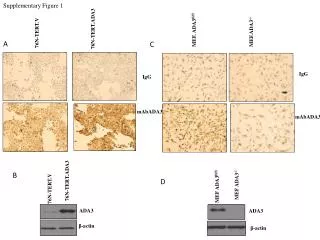
Supplementary Figure1. Characterization of ADA3 monoclonal antibody specificity for IHC staining. (A) IHC staining of vector and flag-tagged ADA3 overexpressing immortalized normal mammary epithelial cells (76N-TERT), using ADA3 mAb and mouse IgG (control). (B) Western blotting of 76N-TERT cells transfected with vector or hADA3. (C) IHC staining of ADA3fl/fl and knock out MEFs. (D) Western blotting of ADA3fl/fl and ADA3-/- MEFs