Download
1 / 81
810 likes | 984 Vues
Part 2 Management of Patients With Oral and Esophageal Disorders. 2ed Years Student, 2ed Semester Miss Iman shaweesh January 2008. Disorders of the Teeth DENTAL PLAQUE AND CARIES.

E N D
Part 2Management of PatientsWith Oral andEsophageal Disorders 2ed Years Student, 2ed Semester Miss Iman shaweesh January 2008
Disorders of the TeethDENTAL PLAQUE AND CARIES • Tooth decay is an erosive process that begins with the action of bacteria on fermentable carbohydrates in the mouth, which produces acids that dissolve tooth enamel. The extent of damage to the teeth depends on the following:
The presence of dental plaque • The strength of the acids and the ability of the saliva to neutralize them • The length of time the acids are in contact with the teeth • •The susceptibility of the teeth to decay
Prevention • Measures used to prevent and control dental caries include practicing effective mouth care, reducing the intake of starches and • sugars (refined carbohydrates), applying fluoride to the teeth or • drinking fluoridated water, refraining from smoking, controlling diabetes, and using pit and fissure sealants
Gerontologic Considerations • Many medications taken by the elderly cause dry mouth, which is uncomfortable, impairs communication, and increases the risk of oral infection. These medications include the following: • Diuretics • Antihypertensive medications • Anti-inflammatory agents • Antidepressant medications
Gerontologic Considerations • Poor dentition can exacerbate problems of aging, such as • Decreased food intake • Loss of appetite • Social isolation • Increased susceptibility to systemic infection (from periodontal disease) • Trauma to the oral cavity secondary to thinner, less vascular oral mucous membranes
DENTOALVEOLAR ABSCESSOR PERIAPICAL ABSCESS • more commonly referred to as an abscessed, involves the collection of pus in the apical dental periosteum (fibrous membrane supporting the tooth structure) and the tissue surrounding the apex of the tooth (where it is suspended in the jaw bone). The abscess has two forms: acute and chronic. Acute periapical abscess is usually secondary to a suppurative pulpitis (a pus-producing inflammation of the dental pulp)
Clinical Manifestations • The abscess produces a dull, gnawing, continuous pain, often with a surrounding cellulitis and edema of the adjacent facial structures, and mobility of the involved tooth. The gum opposite the apex of the tooth is usually swollen on the cheek side. Swelling and cellulitis of the facial structures may make it difficult for the patient to open the mouth.
Management • In the early stages of an infection, a dentist or dental surgeon • may perform a needle aspiration or drill an opening into the pulp chamber to relieve tension and pain and to provide drainage. • After the inflammatory reaction has subsided, the tooth may be extracted or root canal therapy performed. Antibiotics may be prescribed.
Nursing Management • The nurse assesses the patient for bleeding after treatment and instructs the patient to use a warm saline or warm water mouth rinse to keep the area clean. • The patient is also instructed to take antibiotics and analgesics as prescribed, • to advance from a liquid diet to a soft diet as tolerated, and to keep follow-up appointments.
Disorders of the Jaw Temporomandibular disorders are categorized as follows (National Oral Health Information) • • Myofascial pain—a discomfort in the muscles controlling jaw function and in neck and shoulder muscles • • Internal derangement of the joint—a dislocated jaw, a displaced disc, or an injured condyle • • Degenerative joint disease—rheumatoid arthritis or osteoarthritis in the jaw joint
Disorders of the Salivary Glands • Parotitis (inflammation of the parotid gland) is the most common inflammatory condition of the salivary glands, although inflammation can occur in the other salivary glands as well. Mumps (epidemic parotitis), a communicable disease caused by viral infection and most commonly affecting children, is an inflammation of a salivary gland, usually the parotid.
SIALADENITIS • (inflammation of the salivary glands) may be caused by dehydration, radiation therapy, stress, malnutrition, salivary gland calculi (stones), or improper oral hygiene. The inflammation is associated with infection by S. aureus, Streptococcus viridans, or pneumococcus.
SALIVARY CALCULUS (SIALOLITHIASIS) • Sialolithiasis, or salivary calculi (stones), usually occurs in the submandibular gland. Salivary gland ultrasonography or sialography (x-ray studies filmed after the injection of a radiopaque substance into the duct) may be required to demonstrate obstruction of the duct by stenosis. Salivary calculi are formed mainly from calcium phosphate.
Cancer of the Oral Cavity • Cancers of the oral cavity, which can occur in any part of the mouth or throat, are curable if discovered early. These cancers are associated with the use of alcohol and tobacco. • Cancer of the oral cavity accounts for less than 2% of all cancer deaths in the United States. Men are afflicted more often than women.
Pathophysiology • Malignancies of the oral cavity are usually squamous cell cancers. Any area of the oropharynx can be a site for malignant growths, but the lips, the lateral aspects of the tongue, and the floor of the mouth are most commonly affected.
Clinical Manifestations • Many oral cancers produce few or no symptoms in the early stages. Later, the most frequent symptom is a painless sore or mass that will not heal. A typical lesion in oral cancer is a painless indurated (hardened) ulcer with raised edges. Tissue from any ulcer of the oral cavity that does not heal in 2 weeks should be examined through biopsy.
Medical Management • Surgical resection, radiation therapy, chemotherapy, or a combination of these therapies may be effective. In cancer of the lip, small lesions are usually excised liberally; larger lesions involving more than one third of the lip may be more appropriately treated by radiation therapy because of superior cosmetic results.
If the cancer has spread to the lymph nodes, the surgeon may perform a neck dissection. Surgical treatments leave a less functional tongue; surgical procedures include hemiglossectomy (surgical removal of half of the tongue) and total glossectomy (removal of the tongue). • Often cancer of the oral cavity has metastasized through the extensive lymphatic channel in the neck region
Neck Dissection • Malignancies of the head and neck include those of the oral cavity, oropharynx, hypopharynx, nasopharynx, nasal cavity,paranasal sinus, and larynx (Fig) • These cancers account for fewer than 5% of all cancers. Depending on the location and stage, treatment may consist of radiation therapy, chemotherapy, surg or a combination of these modalities.
A radical neck dissection involves removal of all cervical lymph nodes from the mandible to the clavicle and removal of the sternocleidomastoid muscle, internal jugular vein, and spinal accessory muscle on one side of the neck.
Group Discussion • Nursing Management • NURSING PROCESS: THE PATIENT WITH CONDITIONS OF THE ORAL CAVITY • Neck Dissection • NURSING PROCESS: THE PATIENT UNDERGOING A NECK DISSECTION
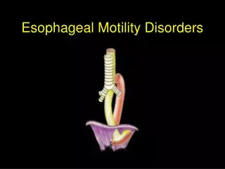
Disorders of the Esophagus • The esophagus is a mucus-lined, muscular tube that carries food from the mouth to the stomach. It begins at the base of the pharynx and ends about 4 cm below the diaphragm. Its ability to transport food and fluid is facilitated by two sphincters. The upper esophageal sphincter, also called the hypopharyngeal sphincter, is located at thejunction of the pharynx and the esophagus. The lower esophageal sphincter, also called the gastroesophageal sphincter, is located at the junction of the esophagus and the stomach.
Dysphagia • (difficulty swallowing) is the most common symptom of esophageal disease. This symptom may vary from an uncomfortable feeling that a bolus of food is caught in the upper esophagus (before it eventually passes into the stomach) to acute pain on swallowing (odynophagia).
Achalasia • is absent or ineffective peristalsis of the distal esophagus accompanied by failure of the esophageal sphincter to relax in response to swallowing. Narrowing of the esophagus just above the stomach results in a gradually increasing dilation of the esophagus in the upper chest. Achalasia may progress slowly and occurs most often in people 40 years of age or older.
Clinical Manifestations • The primary symptom of achalasia is difficulty in swallowing both liquids and solids. The patient has a sensation of food sticking in the lower portion of the esophagus. As the condition progresses, food is commonly regurgitated, either spontaneously or intentionally by the patient to relieve the discomfort produced by prolonged distention of the esophagus by food that will not pass into the stomach. The patient may also complain of chest pain and heartburn (pyrosis).
Assessment and Diagnostic Findings • X-ray studies show esophageal dilation above the narrowing at the gastroesophageal junction. Barium swallow, computed tomography • (CT) of the esophagus, and endoscopy may be used for diagnosis; however, the diagnosis is confirmed by manometry, a process in which the esophageal pressure is measured by a radiologist or gastroenterologist.
Management • The patient should be instructed to eat slowly and to drink fluids with meals. As a temporary measure, calcium channel blockers and nitrates have been used to decrease esophageal pressure and improve swallowing. • Achalasia may be treated conservatively by pneumatic dilation to stretch the narrowed area of the esophagus (Fig. 35-6). Pneumatic dilation has a high success rate.
DIFFUSE SPASM • spasm is a motor disorder of the esophagus. The cause is unknown, but stressful situations can produce contractions of the esophagus. It is more common in women and usually manifests in middle age. • characterized by difficulty or pain on swallowing (dysphagia, odynophagia) and by chest pain similar to that of coronary artery spasm.
Assessment and Diagnostic Findings • Esophageal manometry, which measures the motility of the esophagus and the pressure within the esophagus, indicate that simultaneous contractions of the esophagus occur irregularly. Diagnostic x-ray studies after ingestion of barium show separate areas of spasm.
Management • Conservative therapy includes administration of sedatives and long-acting nitrates to relieve pain. Calcium channel blockers have also been used to manage diffuse spasm. Small, frequent feedings and a soft diet are usually recommended to decrease the esophageal pressure and irritation that lead to spasm.
HIATAL HERNIA • The esophagus enters the abdomen through an opening in the diaphragm and empties at its lower end into the upper part of the stomach. Normally, the opening in the diaphragm encircles the esophagus tightly, and the stomach lies completely within the abdomen. In a condition known as hiatus (or hiatal) hernia, the opening in the diaphragm through which the esophagus passes becomes enlarged, and part of the upper stomach tends to move up into the lower portion of the thorax.
There are two types of hiatal hernias: sliding and paraesophageal • Sliding, or type I, hiatal hernia occurs when the upper stomach and the gastroesophageal junction (GEJ) are displaced upward and slide in and out of the thorax (Fig. 35-8A). About 90% of patients with esophageal hiatal hernia have a sliding hernia. • A paraesophageal hernia occurs when all or part of the stomach pushes through the diaphragm beside the esophagus
Clinical Manifestations • may have heartburn, regurgitation, and dysphagia, but at least 50% of patients are asymptomatic. Sliding hiatal hernia is often implicated in reflux. The patient with a paraesophageal hernia usually feels a sense of fullness after eating or may be asymptomatic.
Assessment and Diagnostic Findings • Diagnosis is confirmed by x-ray studies, barium swallow, and fluoroscopy.
Management • Management for an axial hernia includes frequent, small feedings that can pass easily through the esophagus. The patient is advisednot to recline for 1 hour after eating, to prevent reflux or movement of the hernia, and to elevate the head of the bed on 4- to 8-inch (10- to 20-cm) blocks to prevent the hernia from sliding upward. Surgery is indicated in about 15% of patients.
Management • Medical and surgical management of a paraesophageal hernia is similar to that for gastroesophageal reflux; however, paraesophageal hernias may require emergency surgery to correct torsion (twisting) of the stomach or other body organ that leads to restriction of blood flow to that area.
DIVERTICULUM • A diverticulum is an outpouching of mucosa and submucosa that protrudes through a weak portion of the musculature. Diverticula may occur in one of the three areas of the esophagus—the pharyngoesophageal or upper area of the esophagus, the midesophageal area, or the epiphrenic or lower area of the esophagus— or they may occur along the border of the esophagus intramurally.
The most common type of diverticulum, which is found three times more frequently in men than in women, is Zenker’s diverticulum (also known as pharyngoesophageal pulsion diverticulum or a pharyngeal pouch). It occurs posteriorly through the cricopharyngeal muscle in the midline of the neck. It is usually seen in people older than 60 years of age.
Clinical Manifestations • include difficulty swallowing, fullness in the neck, belching, regurgitation of undigested food, and gurglingnoises after eating. • The diverticulum, or pouch, becomes filled with food or liquid. When the patient assumes a recumbent position, undigested food is regurgitated, and coughing may be caused by irritation of the trachea. • Halitosis and a sour taste in the mouth are also common because of the decomposition of food retained in the diverticulum.
Assessment and Diagnostic Findings • A barium swallow may be performed to determine the exact nature and location of a diverticulum. • Manometric studies are often performed for patients with epiphrenic diverticula to rule out a motor disorder. • Esophagoscopy usually is contraindicated because of the danger of perforation of the diverticulum,