Download
1 / 33
330 likes | 510 Vues
Anatomy & Physiology of Mechanical Digestion. ANATOMY & PHYSIOLOGY 13-14. Avian Digestive Tract. Ruminates. Monogastric Digestive System. Digestive System Divisions. Alimentary Canal (Gastrointestinal Tract) These are all the structures that food passes through during digestion

E N D
Anatomy & Physiology of Mechanical Digestion ANATOMY & PHYSIOLOGY 13-14
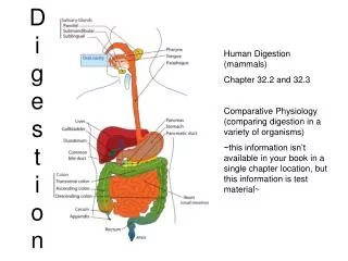
Digestive System Divisions • Alimentary Canal (Gastrointestinal Tract) • These are all the structures that food passes through during digestion • One, long, tube open at both ends • Starting with the mouth, ending with the anus • Accessory Organs • These are the structures that aid in digestion of food, but in which no food passes • Liver, gallbladder, pancreas, salivary glands
Alimentary canal Accessory Organs
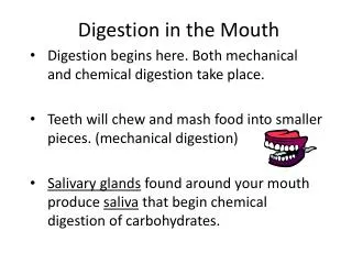
Dentition • Function: Mastication (chewing) • Diphyodont = two sets (20 primary/32 permanent) • Forms: • Incisors (8) Cutting and shearing • Canines (4) Gripping and tearing • Premolars (8) Crushing • Molars (12) Grinding
Palates • Palatum Durum (hard palate): Upper, anterior roof of the mouth, covered with rugae (folds) to allow backwards movement of food • Palatum Velum (i.e. “Soft Palate”): Upper, posterior roof of the mouth. Closes off access to nasopharynx during swallowing
Anatomy of Tongue • Tongue: Muscle that mixes food with saliva, pushes food into the oropharynx, and contains gustatory papillae (taste receptors) • Lingual frenulum: Fold of mucous membrane that attaches the tongue to the floor of the mouth
Tonsils • Lymphoid tissue = first line of immune defense for aerodigestive tract • Largest pre-puberty; atrophy after puberty
Deglutition and Pharyngeal-Esophageal Anatomy • Deglutition = swallowing • Food enters into the oropharynx, then passes into the laryngopharynx • Closure of the epiglottis allows passage of food into the esophagusand prevents aspiration (particulates in lungs)
Peristalsis • Alternating contractions between circular and longitudinal muscle of the pharynx and esophagus • Physically separates food into small spheres (bolus) and moves it through the esophagus
STOMACH • Initial site of protein hydrolysis/digestion • Primary site of mechanical digestion via rumination • Absorption of water and alcohol
Cardiac Sphincter(antrumcardiacum) • Food enters the stomach through this muscle via the esophagus • Accidental opening of this structure may lead to Gastro-Esophageal Reflux Disorder (GERD)
Fundus • Means “bottom” in Latin but is the left anterior curvature of the stomach • Stores food for appx. 1 hour • Digestive gases collect here
Anatomy of Stomach Body • Greater and lesser curvature • Gastric canal – can hold appx. 1 gallon of food • Rugae (increase surface area)
Pyloric Antrum & Sphincter • Muscular terminus of stomach • Involved in rumination of food • Food exits the gastric canal via passage through the pyloric sphincter
DUODENUM • First section of small intestine • Drastic rise in pH due to addition of bile salts • Receives secretions of pancreas
Peptic Ulcers • Most common site of ulcers is 5cm distal to pyloric spincter • Only 4% of ulcers are stomach • Caused by actions of Heliobacter pylori • Exacerbated by stress & diet
SMALL INTESTINE • Jejunum (8ft) • Ileum (12ft) • Site of absorption and chemical digestion
VILLI • Small (1mm) projections of small intestine that drastically increase surface area of small intestine • Cells lining villi die and are consumed! • Contain capillaries for transport of material • Contain lacteals to transport fats
Colonic Form and Function • 1.5m divided into • Ascending • Transverse • Descending • Sigmoid • Involved in conduction of solid waste • Reabsorption of water via standing gradient osmosis
Anal Sphincter • Muscular bands that control expulsion of feces • Regulated via recto-anal inhibitory reflex (RAIR) • Flatuaria is a loss of control over RAIR