Download
1 / 144
1.7k likes | 3k Vues
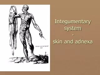
Ocular Adnexa and Lacrimal System. Sometimes, just being around is enough. Introduction. The ocular adnexa includes the structures situated in and around the eyeball This unit will consider: Eyebrows Eyelid structure Conjunctiva Lacrimal system Secretory system Drainage system.

E N D
Ocular Adnexa and Lacrimal System Sometimes, just being around is enough
Introduction • The ocular adnexa includes the structures situated in and around the eyeball • This unit will consider: • Eyebrows • Eyelid structure • Conjunctiva • Lacrimal system • Secretory system • Drainage system
Eyebrows • The eyebrows consist of thick skin covered by short, prominent hairs extending across the superior orbital margin, usually arching slightly but sometimes just running horizontally • Usually, in women the eyebrows run above the orbital margin, and in men they are located along the margin • The muscles in the forehead—the frontalis, procerus, corrugator superciliaris, and the orbicularis oculi—produce eyebrow movement, an important component of facial expression • The frontalis muscle has its origin high on the scalp and inserts into the connective tissue near the superior orbital rim—the fibers run vertically and raise the eyebrow, causing a look of surprise or attention
Eyebrows 2 • The corrugator originates on the frontal bone and inserts into the skin superior to the medial eyebrow—it is the muscle of trouble or concentration, and its fibers are oriented obliquely—it moves the brow, toward the nose, creating vertical furrows between the brows’ • The procerus, the muscle of menace or agression, originates on the nasal bone and inserts into the medial side of the frontalis—it pulls the medial part of the eyebrow inferiorly and produces horizontal furrows over the bridge of the nose • The orbicularis oculi (considered later in this unit) lowers the entire brow • The fibers of these four muscles blend with one another and are difficult to separate—all are innervated by the facial nerve, cranial nerve VII
Eyelids: Introduction • The eyelids or palbeprae, are folds of skin and tissue that, when closed, cover the globe • The eyelids have four major functions • Cover the globe for protection • Move the tears toward the drainage at the medial canthus on closure • Spread the tear film over the anterior surface of the cornea upon opening • Contains glands that produce some of the tear film layers • On closure the upper eyelid moves down to cover the cornea, whereas the lower lid rises only slightly—when the eyes are gently closed, the eyelids should cover the entire cornea
Lagophthalmos • Lagophthalmos refers to the incomplete closure of the eyelids • Its cause may be physiologic, mechanical (e.g., scarring), or paralytic • Lagophthalmos is most evident during sleep, when drying of the inferior cornea may occur • Scratchy, irritated eyes are evident on awakening, and punctate keratitis, sometimes called exposure keratitis, may result • Clinical assessment of the inferior cornea shows varying degrees of epithelial disruption, seen as punctate staining with fluorescein dye
Palpebral Fissure • The palpebral fissure is the area between the two open lids—the upper lid usually just covers the upper limbus, when one is looking straight ahead with open eyes, while the lower lid position is more variable, usually being within 1 mm of the lower limbus • The upper and lower lids meet at the corners of the palpebral fissure in the lateral and medial canthi • The lateral canthus is located about 5 to 7 mm medial to the lateral orbital margin and lies directly on the globe • The medial canthus is at the medial orbital margin, but is separated from the globe by the lacrimal lake, a small pool of tears
Palpebral Fissure 2 • At the floor of the lake is the plica semilunaris, a crescent-shaped fold of conjunctiva that is located in the medial canthus and serves to allow for lateral movement of the eye without stretching the bulbar conjunctiva • The caruncle is a small, pink mass of modified skin/conjunctiva located just medial to the plica • It is covered with epithelium that contains goblet cells and fine hairs and their associated sweat and sebaceous glands
Eyelid Topography • The upper eyelid extends to the eyebrow and is divided into the tarsal and orbital (or preseptal) parts • The tarsal portion lies closest to the lid margin, rests on the globe, and contains the tarsal plate—the skin is thin, and the underlying loose connective tissue without adipose tissue • The orbital portion extends from the tarsus to the eyebrow, and a furrow, the superior palpebral sulcus, separates the tarsal portion from the orbital portion • This sulcus separates the pretarsal skin that is tightly adherent to the underlying tissue, from the preseptal skin, which is only loosely connected to the underlying tissue, which contains a cushion of fat
Eyelid Topography 2 • In the lower eyelid the inferior palpebral sulcus, which separates the eyelid into tarsal and orbital parts, is often not very distinct • The tarsal portion rests against the globe, and the orbital portion extends from the lower border of the tarsus onto the cheek, extending just past the inferior orbital margin to the nasojugal and malar sulci (another name for the zygomatic bone is the malar bone) • These furrows occur at the attachment of the skin to the underlying connective tissue and becomes more prominent with age
Eyelid Margin • The eyelid margin rests against the globe and contains the eyelashes and the pores of the meibomian glands • The cilia (eyelashes) are arranged at the lid margin in a double or triple row, with about 150 in the upper lid and about 75 in the lower • The lashes curl upward on the upper and downward on the lower lid, so they do not completely interlace • Each lash is replaced about every 5 months, and replacement lashes grow to full size in about 10 weeks • The eyelashes are richly supplied with nerves, causing them to be sensitive to even the slightest unexpected touch, which will elicit a protective response—a blink
Abnormalities Affecting the Cilia • Various epithelial diseases can cause madarosis (loss of eyelashes) or trichiasis (misdirection of eyelashes), in which the eyelashes grow toward rather than away from the palpebral fissure • Cilial contact with the cornea can cause irritation and painful abrasions that can become ulcerated • The problem lashes can be removed by epilation
Eyelid Margin • Posterior to the cilia are the pores of the meibomian glands, and the transition from the skin to the conjunctiva, the mucocutaneous junction occurs just posterior to these openings • A groove called the gray line runs along the eyelid margin between the cilia insertions and the pores of the meibomian glands—this line is the location of a surgical plane that divides the eyelid into anterior and posterior portions—it is gray due to an absence of blood supply • The eyelid margin can be divided into two parts: the medial one sixth is the lacrimal portion, and the lateral five sixths is the ciliaryportion
Eyelid Margin 3 • The division occurs at the lacrimal papilla, a small elevation containing the lacrimal punctum, the opening that carries the tears into the nasolacrimal drainage system • Usually, no cilia or meibomian pores are found medial to the punctum, that is, in the lacrimal portion of the lid margin
Epicanthus • Epicanthus is a vertical fold of skin at the nasal canthus, arising in the medial area of the upper eyelid and terminating in the nasal canthal area • It is common in the newborn and may look like an esotropia—a parent of a child with epicanthus might worry that the child’s eyes are crossed, however, a cover test will reveal a true esotropia • As the bridge of the nose develops, epicanthus gradually disappears • A form of epicanthus arising from the tarsal fold and extending into the medial canthal area is common in those of Eastern Asian descent
Eyelid Structures • In this section the following will be considered: • Orbicularis oculi muscle • Superior palpebral levator muscle • Retractor of the lower eyelid • Tarsal muscle (of Müller) • Tarsal plate • Palpebral ligaments • Glands of the eyelid
Orbicularis Oculi Muscle • The elliptical orbicularis oculi muscle acts as a sphincter for the palpebral fissure—its striated fibers extend from the eyelid margin to overlap onto the orbital margin and bones of the brow, temple and cheek • It can be divided into two parts: palpebral and orbital • The palpebral portion of the orbicularis oculi lies in the area of the eyelid that rests on the globe and is the portion of the muscle closest to the eyelid margin • The palpebral part can be further divided into preseptal and pretarsal portions for those parts of the muscle in front of the tarsal plate or the orbital septum
Orbicularis Oculi Muscle 2 • The muscle fibers of the palpebral part form upper and lower semicircles that run from the medial orbital margin and the medial palpebral ligament to the lateral palpebral raphe, where the superior and inferior fibers interdigitate with one another • The lateral palpebral raphe lies over the lateral palpebral ligament • Some fibers arise from deeper origins on the posterior lacrimal crest—this section of the palpebral part of the orbicularis, the muscle of Horner or lacrimal part (pars lacrimalis), surrounds the lacrimal canaliculi
Orbicularis Muscle 3 • Therefore, contraction of the orbicularis assists in moving tears through the canaliculi into the nasolacrimal drainage system • The muscle of Riolan, or ciliary part (pars ciliaris), lies near the lid margin on both side of the meibomian gland openings • Its function is to keep the lid margins close to the eyeball
Ectropion and Entropion • Eversion (a turning away) of the eyelid margin is called ectropion, the common cause of which is loss of muscle tone, a normal part of the aging process • When the lid margin falls away from the globe, the lacrimal punctum is no longer in position to drain the trears from the marginal tear strip (meniscus) in the medial canthus • Epiphora, an overflowing of tears onto the cheek, may occur, causing maceration of the skin in this area • Inversion (a turning inward) of the eyelid margin is called entropion and may result from spasm of the orbicularis • This inward turning causes the eyelashes to touch the cornea and can cause corneal abrasion
Ectropion and Entropion 2 • Scarring of the lid after trauma or disease may cause entropion • Both ectropion and entropion are more common in the lower lid and can be corrected surgically
Orbicularis Muscle 4 • The orbital portion of the orbicularis is attached superiorly to the orbital margin, medial to the supraorbital notch • The fibers encircle the area outer to the palpebral portion and attach inferiorly to the orbital margin, medial to the infraorbital foramen • These concentric circular fibers extend throughout the rest of the lid and over the orbital rim • The orbicularis oculi is innervated by cranial nerve VII (the facial nerve) • Contraction of the palpebral portion closes the eyelid gently, and the palpebral orbicularis is the muscle used in an involuntary blink and a voluntary wink—relaxation of the antagonistic levator occurs simultaneously
Orbicularis Muscle 5 • Spontaneous involuntary blinking renews the tear film • A reflex blink is protective and may be triggered by: • A loud noise • Corneal, conjunctival, or cilial touch • The sudden approach of an object (menace reflex) • When the orbital portion of the orbicularis contracts, the eye is closed tightly and the muscles surrounding the lids—the forehead, temple, and cheek—are involved in contraction • Such a tight closure of the eyelid is often a protective mechanism against ocular pain or after injury and is called reflex blepharospasm
Obicularis Muscle 6 • If the lids are closed by a strong muscular contraction, the forces compressing the orbital contents can cause a significant increase in intraocular pressure • The antagonistto the palpebral portion of the orbicularis is the levator muscle • The antagonistto the orbital portion is the frontalis muscle
Superior Palpebral Levator Muscle • The superior palpebral levator muscle, the retractor of the upper eyelid, is located within the orbit above the globe and extends down into the upper lid • It originates on the lesser wing of the sphenoid bone above and in front of the optic foramen, and its sheath blends with the sheath of the superior rectus muscle located below it • As the levator approaches the eyelid from its posterior origin at the orbital apex, a ligament, the superior transverse ligament (Whitnall’s ligament) may act as a fulcrum, changing the anteroposterior direction of the levator to superio-inferior
Superior Palpebral Levator Muscle 2 • As it enters the eyelid the levator spreads out into a broad fan-shaped tendinous extension, the levator aponeurosis • The aponeurosis spreads out into an extensive sheet posterior to the orbital septum, fanning out across its entire width • These tendinous fibers pass through the submuscular connective tissue; the posterior fibers insert into the lower anterior surface of the tarsal plate, and the anterior fibers run between the muscle bundles of the orbicularis to insert primarily into the skin of the eye lid, although some insert into the intermuscular septa of the orbicularis
Superior Palpebral Levator Muscle 3 • This attachment of the fibers from the levator aponeurosis anchors the skin to the underlying tissues in the pretarsal area of the eyelid and creates the palpebral sulcus • The two side extensions of the levator are referred to as horns • The lateral horn helps to support the lacrimal gland by holding it against the orbital roof, creasing and dividing the gland into orbital and palpebral lobes • The lateral horn then attaches to the lateral palpebral ligament and lateral orbital tubercle • The medial horn is attached to the medial palpebral ligament and the medial orbital rim
Superior Palpebral Levator Muscle 4 • Contraction of the levator causes elevation of the eyelid • The connection between the sheaths of the levator and the superior rectus muscle means that when the eye is elevated, the upper eyelid appropriately raises • The levator is innervated by the superior division of the oculomotor nerve, cranial nerve III • The eyelids close by relaxation of the levator and contraction of the orbicularis oculi • The tonic activity of the levator and the relaxation of the orbicularis holds the eyelid open • With a blink, tonic activity of the levator is suspended, and with a burst of activity, the orbicularis rapidly lowers the lid, followed by a cessation of orbicularis activity and resumption of levator tonicity
Retractor of the Lower Lid • The retractor of the lower lid is the capsulopalpebral fascia (lower eyelid aponeurosis), which is an extension from the sheath of the inferior rectus muscle and the suspensory ligament that inserts into the inferior edge of the tarsal plate • This insertion coordinates lid position with globe movement—the lower eyelid is depressed on globe depression, and the lower lid elevates slightly on upward movement of the globe • The capsulopalpebral fascia also fuses with the orbital septum and sends some fibers to insert into the inferior fornix
Tarsal Muscle (of Müller) • The superior tarsal muscle (muscle of Müller) is composed of smooth muscle and originates on the posteroinferior aspect of the levator muscle • These smooth muscle fibers begin to appear within the striated muscle at the point at which the muscle becomes aponeurotic (i.e., turns to tendon) • The superior tarsal muscle inserts on the superior edge of the tarsal plate • Contraction of Müller’s muscle can cause an added 2 mm of lid elevation
Tarsal Muscle (of Müller) • A similar smooth muscle, the inferior tarsal muscle, is found in the lower lid—it arises from the inferior rectus muscle sheath and inserts into the lower conjunctiva and the lower border of the tarsal plate (either into the plate or tissue near it) • Both tarsal muscles are innervated by the sympathetic nervous system • Their contraction widens the palpebral fissure in conditions of fear or surprise
Ptosis • Ptosis is a condition in which the upper eyelid droops or sags • Its cause is weakness or paralysis of the levator or Müller’s muscle—if only Müller’s muscle is affected the ptosis will be less severe • A patient with ptosis may attempt to raise the upper lid using the frontalis muscle, which results in elevation of the eyebrow and wrinkling of the forehead • Nearly all forms can be treated surgically, usually with resection (shortening) of the levator aponeurosis
Tarsal Plate • Each eyelid contain a tarsal plate (tarsus) that gives the lid rigidity and shapes it to the globe • In the upper lid the tarsal plate is about 11 mm high, and the inferior tarsal plate is only 5 mm high • The anterior surface of the tarsus is adjacent to the submuscular connective tissue layer of the lid—the posterior surface is adherent to the palpebral conjunctiva • The orbital border of the tarsal plates is attached to the orbital septum, and the lid border lies at the lid margin • The sides of the tarsal plates are attached to the bony orbital margin by the palpebral or tarsal ligaments
Palpebral Ligaments • The palpebral or tarsal ligaments are bands of dense connective tissue connecting the tarsal plates to the orbital rim and holding the tarsal plates in position against the globe during lid and eye movements • The medial palpebral ligament runs from the medial edge of each tarsal plate to the medial orbital rim, where it divides into two limbs—one limb attaches to the posterior lacrimal crest and the other to the anterior lacrimal crest • Both limbs lie anterior to the orbital septum
Palpebral Ligaments 2 • The lateral palpebral ligament is located posterior to the orbital septum and attaches the lateral edges of the tarsal plates to the lateral orbital margin at the lateral orbital tubercle • Fibrous connections between the lateral palplebral ligament and the check ligament for the lateral rectus muscle allow a slight lateral displacement of the lateral canthus with extreme abduction • The upper borders of both the medial and lateral ligaments are joined to the expansion (aponeurosis) of the levator tendon, and their lower borders are joined to an expansion of the ligament of Lockwood