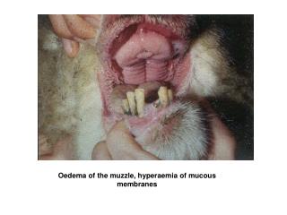
Oedema
Oedema. Extravascular fluid collections can be classified as follows: Exudate extravascular fluid collection that is rich in protein and/or cells fluid appears grossly cloudy Transudate

Oedema
E N D
Presentation Transcript
Extravascular fluid collections can be classified as follows: • Exudate • extravascular fluid collection that is rich in protein and/or cells • fluid appears grossly cloudy • Transudate • extravascular fluid collection that is basically an ultrafiltrate of plasma with little protein and few or no cells. • Fluid appears grossly clear. Effusions into body cavities can be further described as follows: • Serous: a transudate with mainly edema fluid and few cells. • Serosanguinous: an effusion with red blood cells. • Fibrinous (serofibrinous): fibrin strands are derived from a protein-rich exudate. • Purulent: numerous PMN's are present. Also called "empyema" in the pleural space.
Here is an example of fluid collection into a body cavity, or an effusion. This is a right pleural effusion (in a baby). Note the clear, pale yellow appearance of the fluid. This is a serous effusion Pleural Effusion
Here is an example of bilateral pleural effusions. Note that the fluid appears reddish, because there has been hemorrhage into the effusion. This is a serosanguinous effusion. Pleural Effusion
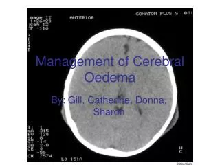
Cerebral Oedema Oedema of the brain is of 3 types: vasogenic, intrstitial ot cytotoxic.It is characterised by raised intracranial pressure (ICP), and results is a swollen brain showing narrowed sulci & flattened gyri.The brain feels soft & fluid drops from the cut surface.
Chronic passive congestion leads to the pathology of "Nutmeg liver". Congestion refers to blood backing up in the liver due usually to right-sided congestive heart failure. With abnormality in the ability of the right heart to pump, blood flow is impaired. This leads to "backwards" heart failure with backup of the venous blood entering the right atrium via the inferior and superior vena cava. A combination of hypoperfusion (lack of blood to cells) and retrograde congestion results. Congestion of Liver
Centrilobular necrosis is evident (review the three zones of a liver lobule), the centrilobular zone receives the most deoxygenated blood and therefore necrosis occurs here first. RBCs are prominent in this zone. The mottled appearance that is observed in centrilobular regions due to this gives rise to the Nutmeg liver description. Congestion of Liver
The alveolar macrophages stain blue with this Prussian Blue iron stain.Such staining indicated iron-laden (haemosideren) macrophagesNote the black pigment in the subpleural connective tissue (coal pigment) Congestion of Lungs
Here are petechial hemorrhages seen on the epicardium of the heart. Petechiae (pinpoint hemorrhages) represent bleeding from small vessels and are classically found when a coagulopathy is due to a low platelet count. They can also appear following sudden hypoxia. Petichiae
The blotchy areas of hemorrhage in the skin are called ecchymoses (singular ecchymosis), or also as areas of purpura. Ecchymoses are larger than petechiae. They can appear with coagulation disorders. Ecchymosis