Download
1 / 154
1.58k likes | 3.25k Vues
Pathology and TCM Treatment of the Herniated Lumbar Disc. East West Healing Center By Dr. Leon Chen www.eastwesthealingcenter.net. D efinition in Western Medicine . Lumbar intervertebral disc injury leads to partial damage to or tears of the annulus fibrosus

E N D
Pathology and TCM Treatment of the Herniated Lumbar Disc East West Healing Center By Dr. Leon Chen www.eastwesthealingcenter.net
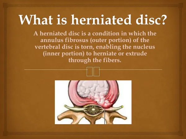
Definition in Western Medicine Lumbar intervertebral disc injury leads to • partial damage to or tears of the annulus fibrosus • protrusion of the nucleus pulposus • compression of the spinal nerve roots • lower back pain, leg pain (including shooting pain) This is called Lumbar Disc Herniation Syndrome.
Definition in Traditional Chinese Medicine (TCM) • Lumbar Disc Herniation Syndrome is called “BiZheng痹症” in Traditional Chinese Medicine (TCM). • The HuangDiNeiJinin 475-221 B.C.(The Yellow Emperor’s Internal Classic) discussed the syndrome of pain in the low back and leg.
Ⅰ Local anatomy The Structure of Vertebral Column The vertebral column in an adult typically consists of 33 vertebrae arranged in five regions: 7 cervical, 12 thoracic, 5 lumbar, and 5 sacral, and 4 coccygeal. The vertebral column is considered to have 26 vertebrae, because 5vertebrae are fused in adults to form the sacrum and 4 vertebrae are fused to form the coccyx.
Curvatures of the Vertebral Column: The vertebral column appears straight from the anterior and posterior position. Laterally, it has three natural curves to balance the body: cervical, thoracic, and lumbar curves. A straight linefrom head to foot should run through the crossing point of each curvature.
Physical Purposes of the Curvatures of the Vertebral Column: • To increase the ability of vertebral column to support weight; and balance the body. • 2) To decrease the concussion to protect the head. • 3) To strengthen the stability of the standing posture. • 4) To spread body weight evenly throughout the vertebrae and discs.
Measurementof lumbarsacralangle can be found by drawing a line along the sacral base (B) and making a horizontal line (A). Normal values lie between 26-57° with a mean of 41°. lumbarsacralangle L3 The lumbar gravity line: the C line from center of L3 body by drawing a vertical line which pass through the anterior lip of the sacral base (S1), if this C line does not surpass 10 mm that is all normal. 41° A B C
Structure of Lumbar Vertebrae: • 1) Lumbar vertebrae have massive and flat bodies, because this shape helps to support more body weight. • 2) Each vertebrae includes the vertebral body (centrum), vertebral foramen, pedicle, lamina,articular facet, articular process, transverse process and spinous process.
椎弓板Lamina 上关节突Superior articular process 棘突 Spinous process 横突Transverse process 椎孔 Vertebral foramen 椎弓根Pedicle 椎体Centrum 上关节突Superior articular process 椎弓根 Pedicle 横突Transverse process 椎体 Centrum 棘突Spinous process 下关节突 Inferior articular facet
Structure of the Intervertebral Disc 1) *Hyaline Cartilage: is the cartilage of the superior and inferior surfaces of the vertebral body. It also forms the top and bottom border of nucleus pulposus. It bears the weight and protects the nucleus pulposus. 2) *Annulus Fibrosus: is a fibrous ring, like a radial tire. It is elastic, embracing and holding the nucleus pulposus, not leting it herniate. 3) *Nucleus Pulposus: is a kind of gelatinous, flexible, semifluid material, located in the center of the annulus fibrosus. Both top and bottom surface are sealed by hyaline cartilage.
Intervertebral Disc 纤维环 髓核 纤维环 椎体 Centrum 透明软骨板 Hyaline Cartilage 纤维环 Annulus Fibrosus Nucleus
Thickness of Intervertebral Discs The Thickness of IV Disc: total: 139mm. Cervical IV disc, 3.85 mm. Thoracic IV disc, 4.03 mm. Lumbar IV disc, 12.7 mm.
Function of Intervertebral Discs : The function of lumbar IV discs is very similar to the intervertebral (IV) discs of the cervical and thoracic vertebra: • To bear the weight of the trunk • To connect to the limbs • To perform normal physical posture and movement. Lumbar IV discs are the most important in the vertebral column.
Function of Intervertebral Discs (2) • Uphold the length of the spinal column and body height. • Connect with adjacent vertebrae. • Bear the weight evenly throughout the vertebral bodies. • Act as a cushion or shock-absorber, protecting the spinal cord and brain. (the major purpose.
Structure of the Spinal Canal 1) The spinal canal is a passage, formed by successive openings in the articulated vertebrae through which the spinal cord and its membranes (epidural space) pass. Also called vertebral canal. 2) The spinal canal is made up of the vertebral foramen, and ligamentum flavum, and posterior longitudinal ligament.
flava ligament posterior longitudinal ligament. Pedicle Superior articular facet
Ⅱ Biomechanicsof the vertebral column • The vertebral column has inner balance and outer balance which helps the body to move in a balanced way. Normally, both inner and outer balance of the vertebral column keeps the body in perfect balance. • Inner balance is formed by discs and facet joints (zygapophysial joints) of vertebrae. • Outer balance is formed by dorsal and ventral muscles.
Anterior longitudinal ligament Posterior longitudinal ligament Centrum Back Muscles Disc Interspinales ligament Spinal Canal abdomen Spinous process Sacrum Abdominal Muscles Pelvis
身体平衡示意图 Balance of body 上肢Upper limbs 颈部 neck 胸腔thorax 腹腔abdominal cavity 横膈midriff 脊柱Vertebral column 骨盆腔pelvis Upper limb: Balance Low limb: Support Vertebral Column: Axis Pelvis: Pivot 下肢 Low limbs
Muscles in the Outer- Balance of Vertebral Column 1) Dorsal muscles: • Psoas Major • Quadratus Lunborum • Sacrospinalis • Latissimus Dorsi • Trapezius • Rhomboideus
2) Ventral muscles: • Serratus posterior inferior • Rectus Abdominis • Transversus Abdominis
T12 L5 Iliaccrest 腰大肌 Psoas Major 腰方肌 Quadratus Lunborum
T6 T12 Thoracolumbar fascia L5 骶棘肌 Sacrospinalis 斜方肌 Trapezius 背阔肌 Latissimusdorsi
C4 T4 8 T11 12 L2 锯齿肌 Serratus posterior inferior 菱形肌 Rhomboideus
Xiphoid process 5 7 pubis 腹直肌 Rectus abdominis 腹横肌 Transversus abdominis
Quadratus Lunborum Latissimus dorsi Transversus abdominis Psoas Major Rectus abdominis
Ⅲ Lumbosacral Plexus Lumbar Plexus 腰丛神经L2~L5 股神经 Femoral N:L2-L4 腰骶神经丛 Lumbosacral Plexus 闭孔神经 Obturator N: L2-L5 Common Peroneal N :L4~S2 Supercficial N Sacral Plexus 骶丛神经S1~S3 坐骨神经 Sciatic N: L4,5;S1,3 Deep N Lateral plantar N Tibial N L4~S3 Medial plantar N
Figure of Lumbosacral Plexus 髂腹下神经 Iliohypogastric N 髂腹沟神经 Ilioinguinal N 腹股沟韧带 生殖股神经 Genitofemoral N Inguinal ligament 股外侧皮神经 Lateral femoral cutaneous N 股神经 Femoral N 阴部神经 Pudendal N 闭孔神经 Obturator N 坐骨神经 Sciatic N
Femoral Nerves • The femoral nerve involves the ventral rami of the spinal nerves of L2-L4. • Distribution: Skin of anterior and medial surfaces of thigh, leg, and foot. • Supplies: the anterior muscles of the thigh (Quadriceps femoris, Sartorius).
Femoral nerve Anterior branches Posterior branches Intermediate cutaneous nerve Medial cutaneous nerve Saphenous nerve
股外皮神经 Lateral femoral cutaneous N Sensory area of Lateral femoral cutaneous N 股神经 Femoral N Saphenous nerve Sensory area of Femoral N Intermediate cutaneous nerve Medial cutaneous nerve
Lateral femoral cutaneous nerve • The lateral femoral cutaneous nerve arises from the spinal nerves of L2 and L3, and travels to innervate the lateral thigh. It supplies the skin on the lateral aspect of the thigh.
L2 L3 L4
Obturator Nerve • The obturator nerve arises from L2-L4 – the ventral rami of the spinal nerves. It supplies the skin on the medial surface of the thigh.
闭孔神经 Obturator N Adductor brevis Adductor longus Adductor magnus Sensory area of Obturator N
Sciatic Nerve • The sciatic nerve is a large nerve that runs down the lower limb. It is the longest single nerve in the body. • The sciatic nerve involves L4, L5 and S1-3 –the spinal nerves of the main sacral plexus. • It includes the common peroneal nerve and the tibial nerve. • It distributes to the skin of the posterior surface of the leg and the sole of the foot.
坐骨大孔 Greater sciatic notch Greater trochanter 坐骨神经 股骨小结节 Lesser trochanter 闭孔神经 Obturator N 闭孔 Obturator foramen Tuberosity of ischium
坐骨神经 Sciatic N Sensory area of Sciatic N 腓总神经 Common Peroneal N 胫神经 Tibial N
Common Peroneal Nerve • It has branches called the superficial and deep peroneal nerves. • The superficial peroneal supplies the muscles of the lateral compartment of the leg. • The deep peroneal supplies the muscles of the anterior compartment of the leg.
腓总神经 Common Peroneal N 腓深神经 Deep nerve 腓浅神经 Superficial nerve 腓浅N感觉支配区 腓深N感觉支配区
Tibial Nerve • The tibial nerve supplies the muscles and skin on the posterior surface of the leg and the sole of the foot. • The tibial nerve gives rise to the sural nerve (which supplies the skin on the back of the leg) and ends on the sole of the foot as the medial and lateral plantar nerves.
腓总神经 Common Peroneal N 胫神经 Tibial N 感觉支配区 胫神经 Tibial N 足底外侧神经 Lateral plantar N 足底内侧神经 Medial plantar N
Location of vertebrae in relation to the conus medullaris • Cervical: Cervical vertebrae: the number of the vertebra plus one corresponds to the number of cervical conus medullaris. • Thoracic: Upper thoracic vertebrae: the number plus two corresponds to the number of the thoracic conus medullaris. Lower thoracic vertebrae: the number plus three corresponds to the number of the thoracic conus medullaris. • Lumbar vertebrae: correspond to the number 1~5 of sacral conus medullaris.
CV2 C-CM8 CV7 T-CM2 TV1 T-CM8 TV6 L-CM1 TV11 L-CM3 TV12 LV1 S-CM1,5 Location of vertebrae in relationship to the conus medullaris
Intervertebral Disc and Nerve Roots LV2 LN3 LV3 LN4 LV4 LN5 LV5 SN1 SV1
3 C C 3 4 5 4 6 5 7 T1 8 2 3 4 5 6 7 T1 8 C5 9 C8 10 11 C7 12 L1 C6 4 S2 L4 L2 5 C6 S2 3 C8 C7 L3 L4 S2 L5 S1 L5 L4 S1 L4 L5 L5
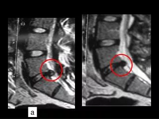
ⅣPatterns of Disc Herniation • Three patterns differentiated by the condition of nucleus pulposus herniation • Five patterns differentiated by the location and direction of nucleus pulposus herniation • Two patterns differentiated by ligament damage • Three patterns differentiated by pathological stages of nucleus pulposus
Three patterns differentiated by the condition of nucleus pulposus herniation • Protrusion or bulging: The annulus fibrosus is not torn but protruding or bulging, compressing the nerve root. • Extrusion: The annulus fibrosus is torn, and the nucleus pulposus herniated to compress the spinal cord or nerve roots. • Sequestration: The annulus pulposus is ruptured, the fragment ofnucleus pulposus has traveled below the posterior longitudinal ligament and herniated into the spinal canal, compressing the spinal cord or nerve root.