Download
1 / 10
100 likes | 398 Vues
Identification of possible biological stains for differential staining of Normal and Malignant tumour tissue. Nor Hayati Othman, Pathology dept, PPSP hayati@kb.usm.my. Potential partner[s]. Chemists Biologists Material engineers. Objectives.

E N D
Identification of possible biological stains for differential staining of Normal and Malignant tumour tissue Nor Hayati Othman, Pathology dept, PPSP hayati@kb.usm.my
Potential partner[s] • Chemists • Biologists • Material engineers
Objectives • To search for suitable natural stains derived from national plants of Malaysia • To determine the differential staining of the new stain on human normal and malignant tumour tissue
Normal Vs cancer cell Normal cell n:c 1:4-8 smooth nuclear membrane no nucleus Malignant cell n:c more hyperchromatic [more basic] nuclear membrane coarse chromatic coarse, stippled mitosis pleomorphic Cytoplasm - ± opaque
Normal colon Malignant colon Normal prostate Malignant prostate Normal cevix Malignant cervix
Normal lung Malignant lung Normal breast Malignant breast Normal thyroid Malignant thyroid
Those are easy cases!! • Subtle changes in cell require further tests • Immunohistochemsitry • We stain the proteins found in cells eg muco-proteins, keratins, vimentins, osteobilins, neurofilaments etc • Every protein has different characteristics and could be stained
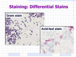
The stain should be • Permanent • Able to differentiate acidic nucleus from basic cytoplasm • Able to pick up loss of desmosomes when normal epithelial cell become malignant • Able to detect changes in nucleus when cells become malignant
How to verify the stains? • 100 Normal and 100 Epithelial Malignant human tissues obtained from the archives of Pathology laboratory. • The ability to differentiate will be scored and statistically analysed
This is possible innovation …but probably time consuming. • Require several trials and errors • Perseverance will be rewarded!