Download
1 / 55
630 likes | 1.6k Vues
Radiology Conference First Trimester Bleeding. Cliff Erickson M.D. & Praveen Reddy, M.D. Albany Medical Center. 1st Trimester. The interval between the last menstrual period (LMP) and 12 weeks gestation. Early Embryogenesis. Ultrasound. Transabdominal less invasive

E N D
Radiology Conference First Trimester Bleeding Cliff Erickson M.D. & Praveen Reddy, M.D. Albany Medical Center
1st Trimester • The interval between the last menstrual period (LMP) and 12 weeks gestation
Ultrasound • Transabdominal • less invasive • wider view of pelvic structures • bladder needs to be full • probe is lower frequency so have lower resolution • Transvaginal • invasive • bladder does not need to be full • higher frequency probe so better resolution • can evaluate in different planes
Definitions • Echogenic - degree to which a structure deflects ultrasound beams. • Anechoic - (or hypoechoic) structures that transmit rather than deflect ultrasound beams and appear black such as water and blood. • Increased echogenecity - appears white like gas, stones, fat and bones.
Case #1 • 22 yo female presents to the ED with c/o vaginal bleeding. The patient denies abdominal pain. • What do you want to know now? • Sexually active, LMP 7 weeks ago • No prior pregnancies • No hx STD/ abnormal gyn exams • What is the next test? • ICON+
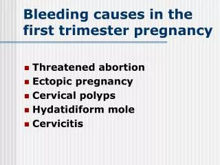
Case #1 Differential Diagnosis • Threatened or spontaneous abortion • Ectopic pregnancy • Normal ovum implantation • occurs around time of expected menses • Subchorionic hemmorhage • Gestational trophoblastic disease • 1:1700 pregnacies
U/S findings of a normal IUP • Identification of a gestational sac with yolk sac or fetal pole confirms IUP • Excludes diagnosis of ectopic pregnancy except if you have a heterotopic pregnancy. Incidence in past was 1/30,000. Now ranges from 1/3,000 to 1/6,000.
Spontaneous Abortion • Loss of pregnancy before 20 weeks/500g • 20-40% of pregnancies • 75% in first 8 weeks • 50-60% due to chromosomal abnormalities • Multiple risk factors • age - • prior SAB • comorbidities • maternal infections
Spontaneous Abortion • Threatened: bleeding, os closed • 50-80% miscarry • Inevitable: bleeding, os open • Incomplete: bleeding, os open, passage of poc • most likely between 6-14 weeks • Complete: bleeding stopped, os closed, poc expelled. • difficult to diagnose in ED
Early Failure • No embryonic cardiac activity with CRL > 5 mm • Embryonic bradycardia relative to CRL. • 100% if CRL < 5 mm and rate is less than 80 bpm or with CRL of 5 - 9 mm and rate < 100 bpm, and CRL 10 - 15 mm with rate < 110 pm. • Gestational sac > 8 mm without a yolk sac • Gestational sac > 16 mm without an embryo • Mean sac diameter minus CRL is less than 5 mm • Poor sac growth. • normally grows 1 mm mean sac diameter per day. • Large yolk sac (> 5.6 mm prior to 10 weeks) • Abnormally large or floppy, amniotic sac
Case # 2 • 25 yo female G3 P1011 with a history of ETOP and PID in the past presents with left sided pelvic pain for two days and intermittent vaginal spotting. • VS P 74 BP 123/72 RR 14 • DDx?
Ectopic Pregnancy: Epidemiology • ~2% of pregnancies • ~40% are missed in first encounter • 4-5x increase in rates over last 20 years • Leading cause of maternal 1st trimester death • second overall to PE • death rate decreased from 35.5/10000 in 1970 to 3.8/10000 in 1989 • nonwhite women have 3.4x increased death rate • teenagers have highest death rate
Ectopic Pregnancy: Risk Factors • History of PID • increases risk 6-7x • each subsequent episode further increases risk • History of prior ectopic pregnancy • Tubal ligation • ectopic until proven otherwise • IUD use • 3-4% of pregnancies with IUD • not type specific • does not continue past removal of IUD
Ectopic Pregnancy: Risk Factors • History of gynecological surgery • ETOP • Prior diagnosis of adhesions • IVF • Chemical ovulation induction • Other assisted reproduction techniques
Ectopic Pregnancy: Risk Factors 50% of patients with an ectopic pregnancy have NO risk factors
Ectopic Pregnancy: History • Menstrual history • amenorrhea 4-12 weeks in 70% cases • 15% of patients report no missed menses • no predictive value • Abdominal pain • “classic” rupture localized, sharp, severe, sudden • 10% may have no pain with EP • 20% with rupture reported shoulder pain • 4% with rupture are pain free
Ectopic Pregnancy: History • Vaginal bleeding • 80% of EP have VB • typically scant/spotting • heavy bleeding suggests SAB but does not rule out ectopic • bleeding usually precedes pain
Ectopic Pregnancy: Physical Exam • Highly variable • Peritoneal signs are present in ~90% of ruptures • Shock, adnexal mass and tenderness “classic” • Fever is rare <2% • Adnexal mass or fullness 65% of patients • 20% on opposite side from EP (corpus luteum cyst) • May have CMT • May have a normal exam
Ectopic Pregnancy: H & P There is no way to exclude ectopic pregnancy on the basis of history and physical examination.
Ectopic Pregnancy Treatment • Medical • methotrexate most common choice • generally useful <6 weeks EGA & <3.5 cm • 5-10% treatment failures • abd pain 3-7 d post administration in 35-55% of pts • Surgical • laparotomy vs laparoscopy • laparoscopic salpingostomy maximizes fertility
Extrauterine Gestation • 95 - 97 % Tubal • 0.5 - 1% Ovary • 0.1 % Cervix
EGA and B-hCG levels • Limited to IUPs only • Doubles every 2 days EGA B-hCG U/S findings 5 weeks 1000 gestational sac 6 weeks 2500 yolk sac 7 weeks 5000 fetal pole 8 weeks 17000 fetal heart beat
Ectopic Pregnancy: Sonographic Findings • Empty nongravid uterus with… • A live embryo in the adnexa • An adnexal mass with a tubal ring (gestational sac) +/- yolk sac • An adnexal mass with no definite tubal ring • Echogenic free fluid • Decidual cyst
Sonographic Findings and Risk of Ectopic T/A Endovaginal Adnexal Embryo 100 100 Mass and large to moderate amount of fluid 100 100 Mass and Fluid 85 78 Any Fluid 71 75 Any Mass 71 69 No mass or Fluid 20 33
Adnexal embryo with heart beat Adnexal mass with yolk sac or embryo Adnexal mass with tubal ring Any adnexal mass, not a simple cyst Decidual cyst 100% 100% 97.8% 96.3% 80% PPV of U/S findings for an ectopic pregnancy