VIROLOGY An Introduction to the Viruses
510 likes | 862 Vues
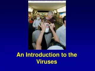
VIROLOGY An Introduction to the Viruses. By Prof. Mohamed I. Bassyouni. General properties of viruses.

VIROLOGY An Introduction to the Viruses
E N D
Presentation Transcript
VIROLOGYAn Introduction to the Viruses By Prof. Mohamed I. Bassyouni
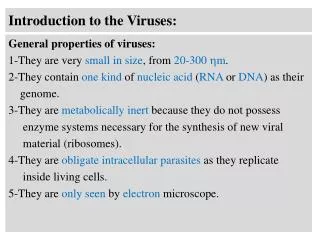
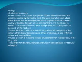
General properties of viruses Viruses are parasites at the genetic level. They are the smallest infectious agents known. They can infect man, animals, insects, plants and bacteria. The following properties distinguish viruses from other organisms: 1- They are very small in size. 2- They contain one kind of nucleic acid (RNA or DNA). 3- They are metabolically inert, as they do not possess ribosomes or protein synthesizing apparatus. 4- They are obligate intracellular parasites. 5- They cannot be grown on artificial culture media and are grown in tissue culture, embryonated eggs or living animals.
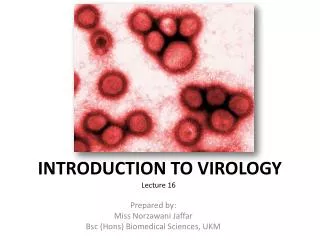
Size of viruses: They vary in size from 20-450 nm. a- They can pass through bacterial filters. b- They require high speeds (ultracentifugation) for their sedmentation 10,000-30,000 rpm ( bacteria require 1,000-3,000 rpm). c- They are only seen by electron microscopy (EM), except poxviruses.
Structures compared From Medical Microbiology, 5th ed., Murray, Rosenthal & Pfaller, Mosby Inc., 2005, Fig. 6-4.
STRUCTURE OF VIRUSES: Each virus particle or virion is composed of a protein coat or capsid and nucleic acid core. The capsid with its enclosed nucleic acid is called the nucleocapsid. Many viruses are naked but some viruses are enveloped. Other internal organs found in some viruses are enzymes, and matrix proteins which are present in enveloped viruses and mediate the interactions between the capsid and envelope.
Viral Capsid: It is composed of small protein subunits called capsomers. The arrangement of the capsomers determines virus symmetry. Functions of the capsids are: 1- It protects the viral genome (DNA or RNA) against inactivation by nucleases. 2- It is responsible for the structural symmetry of virions i.e. icosahedral or helical. 3- It participates in attachment of virions to susceptible cells. 4- Capsid proteins are important antigens that induce antibodies that neutralize virus infectivity and, activate cytotoxic T cells to kill virus-infected cells. 5- Variation in capsid proteins is responsible for the different viral serotypes in non-enveloped viruses.
Icosahedral 20-sided with 12 corners Vary in the number of capsomers Each capsomer may be made of one or several proteins Some are enveloped
complex Fig 6.9a,c
Viral Nucleic Acid (genome) Viruses contain either DNA or RNA but not both. Most DNA viruses are double stranded, while most RNA viruses are single stranded. The nucleic acid may be linear or circular. Some RNA viruses have segmented genome e.g. rotavirus and influenza virus. The molecular weight and type of nucleic acid are specific for each virus group. All viruses have one copy of either genome (haploid) except retroviruses which have two copies (diploid). Viral genomes are used as vectors in gene therapy and in recombinant a virulent virus vector vaccines. Functions of the nucleic acid are: 1- It is the infectious part of the virus; coreless particles are non-infections. 2- It carries the genetic information for (a) Virus replication. (b) Virulence or ability to parasitize cells. (c) Antigenic specificity of the protein coat.
Viral Envelope Many viruses are surrounded by a lipid or lipoprotein, which may be covered by glycoprotein spike-like projections, which attach to host cell receptors during the entry of the virus into the cell, e.g. haemagglutinin (HA) and Neuraminidase (NA) spikes in influenza virus. Due to their lipid content, such viruses are sensitive to ether. Loss of lipids results disruption of virus and loss of infectivity. The envelope may be partially or completely derived from host membranes during release from the cell by budding. The surface proteins, whether the virus capsid proteins or the envelope glycoproteins, are the principal antigens against which the host mounts its immune response. They are also the determinants of type specificity.
Viral Enzymes Some viruses carry enzymes e.g. RNA polymerase, which is present in negative sense RNA viruses to copy their mRNA e.g. orthomyxoviruses and the reverse transcriptase enzyme (RT) present in retroviruses to make a cDNA copy of the viral RNA. Virus Symmetry The arrangement of the capsomers, in the capsid, gives the virus its geometric symmetry. Viruses have three types of symmetry: 1- Cubical symmetry; These viruses resemble a crystal and are called icosahedralviruses e.g. herpsviruses and adenoviruses.
2- Helical symmetry in which the particle is elongated. The capsomers are arranged in a ribbon which is wound in the form of a helix or spiral around the spiral nucleic acid. All human viruses that have helical symmetry are enveloped e.g. influenza virus. 3- Complex symmetry in which the viruses are complicated in structure e.g. poxviruses which are brick shaped with ridges on the external surface. The bacteriophage is another example of complex symmetry. Atypical virus-like agents: 1- Defective viruses are composed of viral nucleic acid and proteins but cannot replicate without a helper virus, which provides the missing function. These usually have a mutation or a deletion of part of their genetic material. During the growth of most human viruses, many more defective than infectious virus particles are produced.
2-Pseudovirionscontain host cell DNA instead of viral DNA within the capsid. They are formed during infection with certain viruses when the host cell DNA is fragmented and pieces are incorporated within the capsid. Pseudovirions can infect cells, but they do not replicate. 3- Prionsare infectious particles that are composed solely of protein. They contain no detectable nucleic acid. They cause slow diseases.
6 steps in bacteriophagereplication • Adsorption– binding of virus to specific molecule on host cell. • Penetration–genome enters host cell. • Replication– viral components produced. • Assembly - viral components assembled. • Maturation– completion of viral formation. • Release– viruses leave cell to infect other cells.
Not all bacteriophageslyse cells • Temperate phages insert their viral DNA into the host chromosome & viral replication stops at there until some later time. • Lysogeny- bacterial chromosome carries phage DNA
Host range • Spectrum of cells a virus can infect • cell has to have a specific structure (receptor) on its surface for viral attachment • cell has to contain all of the enzymes and materials needed to produce new virions • May be one species or many • HIV (only humans) vs rabies (many animals) • May be one tissue or many within a host • Hepatitis (liver) vs polio (intestinal & nerve cells)
Differences between phage and animal virus replication • Animal virus replication is more complex than phage replication because host cells are more complex. • Animal viruses cannot inject their DNA. • Lysogeny for phage, latency for animal viruses
Animal virus replication 1-Adsorption. 2-Penetration/uncoatingof genome. 3-Eclipse. 4-Duplication/synthesis. 5-Assembly. 6-Release.
1- Attachment: Virus and cell are brought into contact by random collision, but attachment is specific and occurs only if the cell membrane contains specific receptors for the virus, e.g. human immunodeficiency virus (HIV) binds to CD4 receptors on immune cells i.e. helper T cells, macrophages and dendritic cells. • 2- Penetration and uncoating: Non-enveloped virions are taken into animal cells by endocytosis where they are uncoated by lysosomal enzymes. Enveloped viruses penetrate the membrane by fusion between the virus envelope and the cell membrane releasing the nucleocapsid into the cell; uncoating my occur at the cell surface e.g. bacteriophages, in the cytoplasm e.g. poliovirus, or in the nucleus e.g. herpesvirus. Uncoating renders viral nucleic acid accessible for transcription.
3- Eclipse: It is the period after penetration during which no infectious virus can be detected inside the host cell. During this phase, the cell is redirected, by the viral nucleic acid (genome), toward synthesizing viral components. The eclipse phase ends with the appearance of virus particles. • 4- Intracellular viral synthesis: It includes synthesis of both viral nucleic acid and proteins. The viral nucleic acid (genome) replicates by using a strand of the parental nucleic acid as a template for the production of progeny DNA or RNA molecules. • 5- Assembly of viral nucleic acid and protein coats to form mature virus particles occurs in the cytoplasm e.g. poliovirus or in the nucleus e.g. herpes viruses.
6- Release : Mature virus particles will accumulate in the cell in enormous numbers and are released by either of two processes. One is by rupturing the cell i.e. cytolysis, which usually occurs with non-enveloped viruses. The other is by a slow process of leaking or budding through the cell membrane, which occurs with enveloped viruses where they acquire lipoprotein envelope during budding from the outer cell membrane ( this is mediated by matrix proteins). Herpes virus acquire their envelope from the nuclear membrane.
Cytopathiceffects- virus-induced damage to cells 1-Changes in size & shape. 2-Cytoplasmic inclusion bodies. 3-Nuclear inclusion bodies. 4-Nells fuse to form multinucleated cells. 5-Cell lyses. 6-Alter DNA. 7-Transform cells into cancerous cells.
How do we grow viruses? Obligate intracellular parasites require appropriate cells to replicate.
Growing animal viruses 1-Live laboratory animals 2-Bird embryos – chicken, duck; intact, self- supporting unit, sterile, self-nourished 3-Cell culture
Other noncellular infectious agents Prions- misfolded proteins, contain no nucleic acid cause spongiform encephalopathies – holes in the brain common in animals scrapie in sheep & goats bovine spongiform encephalopathies (BSE), aka mad cow disease humans – Creutzfeldt-Jakob Disease Viroids- short pieces of RNA, no protein coat only been identified in plants, so far.
Diagnosis of viral diseases - More difficult than other agents - Consider overall clinical picture -Take appropriate sample - Infect cell culture- look for characteristic cytopathic effects - Screen for parts of the virus - Screen for immune response to virus (antibodies)
LABORATORY DIAGNOSIS OF VIRAL INFECTIONS. • 1- Microscopy • * Electron microscope is used to determine virus particles. • * Large viruses may be seen by light microscope e.g. small pox virus. • * Immunoelectron microscope; addition of antibodies causes aggregation of virus particles. • * Immunofluorescent. • * Inclusion bodies can be seen either intranuclear or intracytoplasmic and are helpful in the diagnosis of certain virus infections.
2- Virus Culture • Cell cultures, chicken embryos, or laboratory animals are used for recovery of viruses from clinical samples to establish disease etiology. • The type of cell culture used for virus cultivation depends on the sensitivity of the cell to a particular virus (i.e. viral tropism). • 3- Detection of Viral Antigens • The recognition of hepatitis B antigens in sera of patients, rota virus antigen in feces, rabies virus antigens in brain, Herpes simplex virus in vesicular lesions is achieved by: • * Direct immunofluorescence using specific antibody labeled with fluorescent substance, e.g. diagnosis of rabies in brain tissue of animals. • * Detection of viral antigens in sera of patients by radioimmunoassay or enzyme immunoassay e.g. hepatitis antigens.
4- Nucleic Acid Hybridization • This rapid diagnostic method is based on using fragments of DNA or RNA that are complementary to the viral nucleic acid (molecular probe) labeled with an enzyme or a radioactive substance. This method is highly specific and sensitive. • 5- Polymerase Chain Reaction (PCR) • This technique involves amplification of a short sequence of a specific viral DNA or RNA leading to accumulation of large amounts of that nucleic acid sequence which can be detected easily.
6- Serological Methods • Antiviral antibodies appear 10 days after exposure to infection. In the acute phase of the disease, they are of the IgM class or detection of a 4-fold or greater rise of antibody titer (IgG class). The procedures used are complement fixation, virus neutralization, hemagglutination inhibition, indirect immunofluorescence, radioimmunoassay and enzyme immunoassay.
PATHOGENESIS OF VIRUS INFECTIONS • Viruses should reach a susceptible cell before they can produce disease. Therefore, they should have: • 1- Portal of entry; namely the respiratory tract, gastrointestinal tract (GIT), skin, urogenital tract or conjunctiva. • 2- A pathway through the body; namely the blood, lymphatics or nerves. • 3- A target organ which may be CNS, skin, glands, liver …etc.
Many viral infections are subclinical. The same virus may produce a variety of diseases. The same disease may be produced by a variety of viruses. The outcome of virus infections is determined by the interactions of virus and the host and is influenced by the genetics of each. Viruses may produce local or systemic infections, which may reach the full-blown picture, or more commonly end in subclinical form. Some viruses persist. • 1- Local Infections which occur at the portal of entry with no viraemia e.g. viral influenza and common cold at the mucous membrane of the respiratory tract. Rotavirus infection at the GIT causing diarrhoea. It is characterized by short incubation period, short lasting immunity that is mediated by IgAand interferon.
2- Systemic Infections: after primary replication at the site of entry, the virus travels through the blood or lymphatics causing viraemia, or through the nerves to reach a distant target organ that has specific receptors for the virus e.g. poliovirus, mumps and measles. • Systemic infections are characterized by: long incubation period, long lasting immunity that is mediated by IgG and IgM. Infection can be stopped at the viraemic stage immune mechanisms i.e. neutralizing antibodies. This leads to subclinical or abortive infections. Gamma globulins given to contacts of a case may abort infection if given during the incubation period before viraemic stage. • 3- Persistent viral infections: Sometimes virus persist for a long time in the host in one of the following forms :
a- Chronic infections in which the virus can be continuously detected with no or mild symptoms e.g. hepatitis B and C chronic carriers. • b- Latent infections in which the virus persists hidden most of the time, with periodic reactivation and development of clinical lesions containing the virus e.g. herpes viruses and HIV infections. • c- Slow virus infections the have a very long incubation period of months or years with no clinical symptoms e.g. subacutesclerosingpanencephalitis with incubation period of 2-20 years and is caused by a variant of measles virus and HIV. Other slow infections caused by non-conventional agents called prions e.g. Creutzfeldt-Jakob disease and kuru in man.
Mechanisms that help persistence of virus include: • 1- Integration of a DNA provirus into host cell DNA e.g. retroviruses. • 2- Rapid antigenic variation. • 3- Virus spread from cell to cell without an extracellular phase. • 4- Decreased expression of MHC-1 by the action of viral genes which leads to decreased recognition of virus infected cells by CTLs. • 5- Location within an immunologically sheltered sanctuary e.g. the brain. • 6- Formation of virus-antibody complexes which remain infections. • 7- Immunosuppression as in AIDS.