Download
1 / 58
590 likes | 979 Vues
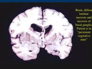
Brain, diffuse laminar necrosis and necrosis of basal ganglia. Patient is in “persistent vegetative state”. Pale infarct, middle cerebral artery. Hemorrhagic infarct, middle cerebral artery. Basal ganglia hemorrhage. Arteriosclerosis due to hypertension.

E N D
Brain, diffuse laminar necrosis and necrosis of basal ganglia. Patient is in “persistent vegetative state”
Fibrinoid necrosis of intracerebral artery secondary to hypertension
Brain, laminar necrosis, focal area
B12 deficiency: myelin loss in lateral and posterior columns
Normal midbrain (left) and Parkinson’s disease (right) Note loss of pigment in substantia nigra
Brain, Perivascular infiltration by lymphocytes in encephalitis, H&E
Brain, Intranuclear inclusion body in neurons in encephalitis, H&E
Brain, Intracytoplasmic inclusion in Purkinje cells in Rabies, H&E
Gray matter: Spongiform encephalopathy in Creutzfeldt-Jakob disease, H&E
Whorling of tumor cells in meningioma
Psammoma bodies in meningioma
Astrocytoma in the temporal lobe
Glioblastoma multiforme; pseudopalisades of tumor cells around necrosis
Cystic astrocytoma of cerebellum
Calcification in oligodendroglioma
Ependymoma in fourth ventricle
Ependymoma; perivascular pseudo-rosettes
Homer Wright rosettes in medulloblastoma
Antony A (dense area) and Antony B (pale area) fibers of schwannoma