CERVICAL TRIANGLES
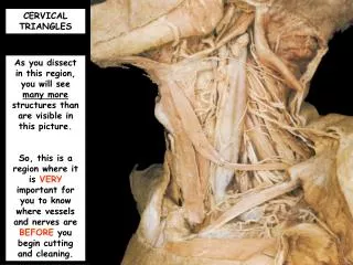
CERVICAL TRIANGLES. As you dissect in this region, you will see many more structures than are visible in this picture. So, this is a region where it is VERY important for you to know where vessels and nerves are BEFORE you begin cutting and cleaning. Superficial temporal v.

CERVICAL TRIANGLES
E N D
Presentation Transcript
CERVICAL TRIANGLES As you dissect in this region, you will see many more structures than are visible in this picture. So, this is a region where it is VERY important for you to know where vessels and nerves are BEFORE you begin cutting and cleaning.
Superficial temporal v. Review the anatomy of the VEINS of this region. Facial v. Internal jugular v. External jugular v. Cephalic v. Axillary v. Subclavian v. Brachiocephalic v. Anterior jugular v. Superior vena cava
Review the anatomy of the ARTERIES of this region. Occipital a. Superficial temporal a. Transverse facial a. Internal carotid a. External carotid a. Superficial cervical a. Facial a. Suprascapular a. Anterior circumflex humeral a. Superior thyroid a. Axillary a. Thoracoacromial a. Thyrocervical trunk Lateral thoracic a. Subclavian a.
AND, where are those all important phrenic and vagus nerves that we want to SAVE? To best observe these structures, you need to cut or reflect the internal jugular vein which allows you a good view of the common carotid artery and the anterior scalene m..
So, once the internal jugular vein is reflected, you will be able to see these nerves. Internal jugular v. Vagus n. – this nerve literally lies within the carotid sheath. Phrenic n. – this nerve lies on the anterior surface of the anterior scalene m..
CERVICAL TRIANGLE SKIN INCISIONS • Make a VERY, VERY SUPERFICIAL incision in the midline of the neck from the tip of the chin to the suprasternal notch. • Make a VERY, VERY SUPERFICIALincision along the clavicle from its medial end to a point that is about 3 cm. beyond the acromion process. • Make a VERY, VERY SUPERFICIAL incision just anterior to the anterior border of the sternocleidomastoid m.. • Reflect skin laterally.
Reflect the platysmam. superiorly towards the mandible. Be VERY careful to not damage the underlying attached structures.
With the platysmam. reflected, note the vessels that you should observe in the anterior cervical triangle. External jugular vein Anterior jugular veins
Identify the boundaries of the posterior cervical triangle. Anteriorly, it is bounded by the posterior edge of the sternocleidomastoid m.. Posteriorly, it is bounded by the anterior edge of trapezius m.. Inferiorly, it is bounded by the clavicle.
External jugular vein Great auricular nerve You should be able to identify some of the most superficial structures in this region, such as the largest cutaneous nerve of the cervical plexus, the great auricular nerve (C2, C3). It crosses the surface of the superior part of the sternocleidomastoid muscle and lies adjacent to the external jugular vein (which drains into the subclavian vein).
Transverse cervical nerve Erb’s point The transverse cervical nerve (C2, C3) crosses the sternocleidomastoid m. transversely. These cervical nerves emerge from a region that is called Erb’s point.
Find the spinal accessory nerve (CN XI) passing deep to the sternocleidomastoid muscle. It crosses the posterior triangle. This nerve is responsible for innervating the sternocleidomastoid and trapezius mm.. Recall that you make mention of this nerve when you teach the cranial nerves.
To examine the structures at the base of the posterior triangle, it may be necessary to cut through the clavicle just lateral to the attachment of the sternocleidomastoid muscle. Only perform this bone cut if you have been told to do so!!!!
Superior belly of the omohyoid m. The two bellies of the omohyoid m. are connected by an intermuscular tendon, that is held by connective tissue to the anterior surface of the internal jugular vein to prevent the vein from collapsing under negative pressure. Inferior belly of the omohyoid m.
Anterior scalene m. Suprascapular a. Transverse cervical a. At this point you should clearly see the anterior scalene m. and two arteries that overlie it. Even though we do not typically show these two arteries during lab, we do show the vessel from which they originate, the thyrocervical trunk.
Phrenic n. Anterior scalene m. As you clean the anterior scalene m.,PLEASE do NOT damage the phrenic n. which should be found running vertically along its medial border. The fibers of this nerve are generally derived from C3, 4, and 5. Recall that each phrenic n. innervates one- half of the diaphragm.
Middle scalene m. Splenius capitus m. Levator scapulae m. Besides the anterior scalenem., you should be able to clearly see some of the other “prevertebral muscles” that form the floor of this posterior triangle.
Anterior scalene m. Middle scalene m. Trunks of the brachial plexus Between the anterior scalene and the middle scalenemm., identify and clean the trunks of the brachial plexus.
So, you should now have a posterior cervical triangle with the muscles shown in this picture cleaned of their overlying fascia. Splenius capitus m. Middle scalene m. Levator scapulae m. Anterior scalene m. Posterior scalene m. Inferior belly of the omohyoid m. Trunks of the brachial plexus
Notice some of the nerves that are found in this region. Dorsal scapular nerve that innervates the rhomboid and levator scapulae mm. Long thoracic nerve that innervates the serratus anterior m. Suprascapular nerve that innervates the supraspinatus and infraspinatus mm. Trunks of the brachial plexus
Now, continue your dissection by finding and cleaning the contents of the anterior cervical triangles. Note the boundaries of each of these triangles are the sternocleidomastoid m., the midline of the neck, and the mandible.
Within these triangles is found the hyoid bone at the angle between the floor of the mouth and the superior neck. Hyoid bone
Four ribbon-like (strap-like) muscles are found on each side descending from the hyoid bone or the thyroid cartilage. As a group, they are called the infrahyoid mm.. Recall that their names basically describe their attachments. Superior belly of the omohyoid m. Sternohyoid m.
Thyrohyoid m. Sternothyroid m.
If these infrahyoid muscles are retracted laterally, you can observe some other important structures. Thyroid cartilage Cricoid cartilage First tracheal ring
Cricothyroid membrane Cricothyroid muscles Incisions through the cricothyroid membrane are a way to quickly access the airway inferior to the vocal cords (cricothyrotomy). structures.
http://www.aic.cuhk.edu.hk/web8/dilational_cricothyrotomy.htmhttp://www.aic.cuhk.edu.hk/web8/dilational_cricothyrotomy.htm
Isthmus of thyroid gland Lobes of thyroid gland
Now look at structures contained within the carotid triangle of the anterior cervical triangle. Posterior belly of the digastric m. Superior belly of the omphyoid m. Anterior border of the sternocleidomastoid m.
Submandibular gland. Common carotid a.
This is a better view of that artery with the SCM cut and reflected. Do NOT perform this reflection unless you are told to do so. External carotid a. Internal carotid a. Common carotid a.
However, if you do reflect the sternocleidomastoid m., you should be able to easily view the spinal accessory nerve (CN XI) on its underside.
While you are in this region, you will observe some nerves which are components of the ansa cervicalis which supply the infrahyoid muscles. Superior root of ansa cervicalis Inferior root of ansa cervicalis
If you are told to do so, cut the common facial vein where it joins the internal jugular vein.
You should now be able to get a good view of the hypoglossal nerve (CN XII). It generally lies just medial to the posterior belly of the digastric m. Recall that this is the motor nerve to the tongue.
Look even more superior to see the occipital artery. Occipital a. Hypoglossal nerve (CN XII)
Other structures of interest in this region are the internal jugular vein and the vagus nerve. Superior root of the ansa cervicalis Superior thyroid a. Internal jugular v. Vagus nerve
Submental triangle Submandibular triangle Identify and clean the suprahyoid muscles.
Identify and clean the suprahyoid muscles. Anterior bellies of the digastric mm. Mylohyoid m. Stylohyoid m. Hypoglossal n. Posterior belly of the left digastric m.