Lesson # 18
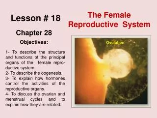
The Fem ale Reproductive System. Lesson # 18. Chapter 28. Objectives:. Ovulation. 1- To describe the structure and functions of the principal organs of the female repro- ductive system. 2- To describe the oogenesis.

Lesson # 18
E N D
Presentation Transcript
The Female Reproductive System Lesson # 18 Chapter 28 Objectives: Ovulation 1- To describe the structure and functions of the principal organs of the female repro-ductive system. 2- To describe the oogenesis. 3- To explain how hormones control the activities of the reproductive organs. 4- To discuss the ovarian and menstrual cycles and to explain how they are related.
The Female Reproductive System Main Reproductive Organs or Gonads Accessory Glands and Organs External Genitalia Duct System Uterine or Fallopian tubes Bartolini’s or greater vestibular glands Uterus Paraurethral glands Vagina Clitoris Labia minora Ovaries Labia mayora
Uterine or Fallopian tube Ovary Uterus Vagina Fornix Paraurethral glands Clitoris Labium majus Bartolini’s or greater vestibular gland Labium minus The Female Reproductive System
Ovarian ligament Suspensory (infundibulopelvic) ligament They perform three main functions: The Ovaries 1- Production of the female gametes or oocytes. 2- Secretion of female sex hormones (estrogens and progestins). 3- Secretion of inhibin (feedback control of pituitary FSH). It attaches the ovary to the uterus. • It attaches the ovary to the pelvic wall and contains ovarian artery, vein and nerves. Ovary Ovary Uterus Broad ligament It is a sheet of peritoneum that flanks the uterus and holds the uterine tube in its superior margin. Broad ligament: Mesovarium It attaches the ovary to the broad ligament.
Ovarian ligament Suspensory (infundibulopelvic) ligament The Ovaries It attaches the ovary to the uterus. The egg develop in their own fluid-filled follicles • It attaches the ovary to the pelvic wall and contains ovarian artery, vein and nerves. It is occupied by major arteries and veins. Medulla • It is where gametes are produced. • Follicle bursting and releasing the egg (ovulation) Cortex (Dense connective tissue) Corpus luteum Tunica albuginea Mature follicle Corpus albicans
Uterine or Fallopian tubes Ampulla Isthmus Infundibulum Fimbriae The Uterine Tubes Ovary Ovary Uterus
Histology of the Uterine Tubes Microvilli of mucin-secretingcells Isthmus Ampulla Infundibulum Fimbriae Uterus Regions of the uterinetubes, posterior view Simple Columnar epithelium Cilia Smooth muscle LM 122 Isthmus Epithelial surface SEM 4000 A sectional view of the isthmus A colorized SEM of the ciliatedlining of the uterine tube
Functions: 1- They receive and transport the secondary oocytes and the fertilized ova to the uterus. Oocytes are transported by a combination of cilliary movements and peristaltic contractions of the wall of the tube. 2- While within the tube, the oocyte may encounter sperm and become fertilized prior to entering the uterus. Ovulation Implantation DAYS 7-10
Endometrium Myometrium Perimetrium Internal os Isthmus Cervical canal External os The Uterus 1- It receives, protects, and nourishes the fertilized egg. Functions: 2- It is the site of the menstruation, development of the embryo and fetus during pregnancy; and of labor. 3- It is a passageway for the sperm. Fundus Body Cervix Vagina
Endometrium Myometrium Perimetrium The Uterine Wall Functional zone Basilar zone Supply functional zone Supply basilar zone Uterine glands Simple columnar epithelium Straight artery Spiral artery Radial artery Uterine artery Arcuate artery
Endometrium Basilarzone Uterineglands Functionalzone Simplecolumnarepithelium Myometrium Uterinecavity Uterine wall LM 32 The basic histological structure of theuterine wall
Bladder Vagina Rectum Urethra Vaginal orifice The Vagina Fornix
External os Histology of the Vagina Stratifiedsquamousepithelium(nonkeratinized) Lumen ofvaginalcanal Bundles of smoothmuscle fibers Bloodvessels Laminapropria Cervix Fornix Rugae Vaginalcanal Greatervestibular gland Vestibule Labia minora The vaginal wall LM 25 Functions: 1- Passageway for the baby, menstrual flow, and sperm. 2- It is the female copulatory organ.
Mons pubis Prepuce Urethral opening Glans or clitoris Hymen (torn) Labia minora Vaginal entrance Labia majora The External Genitalia It is a pad of adipose tissue covering the symphysis pubis.
Lactiferous duct Lactiferous sinus Lobules Suspensory ligament Nipple Areola The Mammary Glands By the end of the six month of pregnancy the mammary glands are fully developed (prolactin hormone produced by the adenohypophysis). They produce milk to nourish the baby. Pectoralis major After birth Pectoral fat pad
Oogenesis Oogonia Diploid SPERMATOGENESIS OOGENESIS MITOSIS Primary First Second Secondary Secondary Diploid oocyte polar oocyte oocyte polar MEIOSIS I Before birth It stops in It stops in metaphase prophase After puberty MEIOSIS I MEIOSIS II Completed Completed Haploid body body MEIOSIS II Before ovulation After ovulation Haploid If fertilization occurs
Between the third and seventh month of fetal life: Oogonia undergo mitosis and produce primary oocytes (diploid). Primary oocytes (diploid) begin MEIOSIS I but it is stopped in prophase I. At birth: Primordial follicles Primary oocytes (diploid) in prophase I of MEIOSIS I. At puberty: Primary follicles FSH triggers the start of the ovarian cycle. Secondary follicles MEIOSIS I is completed to form one secondary oocyte (haploid)and the first polar body. Ovulation Tertiary follicles During reproductive life: Every month one secondary oocyte begins MEIOSIS II that is stopped in metaphase II. Ovulation occurs, and if the secondary oocyte is fertilized, MEIOSIS II is completed to form the ovum and the second polar body.
Comparison between Oogenesis and Spermatogenesis. Like spermatogenesis, oogenesis produces haploid gametes by means of meiosis. Spermatogenesis 1- Males produce sperm continually. 2- Spermatogonia and spermatocytes are produce continually during fertile period of life. Oogenesis 1- It is a cyclic event that releases one egg per month. 2- Oogonia and primary oocyte are produce only before birth. • 3- Oogonia multiply until the fifth month reaching 5 to 6 million in number. Then go into a state of arrested development until shortly before birth 4- Shortly before birth oogonia transform into primary oocyte. 5- Most of primary oocytes undergoes degeneration before the girl is born and during childhood. 6- By puberty, only about 400, 000 remains (even if a woman ovulates every 28 days from 14 to 50, she would ovulate 480 times).
Oogenesis and Sexual Cycle Sexual Cycle Reproductive Cycle The events that recur every month when pregnancy does not intervene. The sequence of events from fertilization to giving birth. • It consists of two interrelated cycles controlled by shifting patterns of hormone secretion: 1- Formation of Primary Follicles 1- Follicular phase 2- Formation of Secondary Follicles • The Ovarian Cycle 3- Formation of Tertiary Follicles (The events in the ovaries) 4- Ovulation 5- Formation of the corpus luteum 2- Luteal phase 6- Degeneration of the corpus luteum Menses • The Uterine (menstrual) Cycle Proliferative phase (The parallel events in the uterus) Secretory phase
The Ovarian Cycle 1- Follicular Phase Follicular fluid First polar body Oocyte at Ovulation Corona radiata Zona pellucida It is a layer of glycoprotein gel secreted by granulosa cells around the oocyte. It is composed of several layers of granulosa cells.
The Ovarian Cycle 2- Luteal Phase Corpus luteum Corpus albicans The corpus luteum produces progesterone, which primary function is to prepare the uterus for pregnancy by stimulating the maturation of the endometrium and the secretion of uterine glands. If fertilization does not occur, progesterone and estrogen levels fall, and the corpus luteum disintegrates and becomes a pale scar called corpus albicans. It also produces moderated amounts of estrogens. It marks the end of the ovarian cycle.
Hormones and the Female Reproductive Cycle FOLLICULAR PHASE LUTEAL PHASE 1- Gonadotropin releasing hormone (GnRH) is produced by the hypothalamus. It stimulates the adenohypophysis to produce gonadotropins: FSH and LH 3- At about day 14, a massive release of LH is produced and ovulation is triggered. 4- The high level of LH that triggers ovulation also promotes the formation of corpus luteum and progesterone secretion. 2- FSH triggers each month the development of one primordial follicle into primary follicles. Primordial Ovulation It is produced by the hypothalamus and stimulates the adenohypophysis to release FSH and LH (Gonadotropins). 1- Gonadotropin releasing hormone (GnRH): Secondary Tertiary Primary It is produced by adenohypophysis and stimulates the development of the follicles. 2- Follicle stimulating hormone (FSH): 3- Luteinizing hormone (LH): It is produced by adenohypophysis and stimulates ovulation. 4- Progesterone: It is produced by the corpus luteum and prepares the uterus for pregnancy.
The Uterine Cycle Functional zone Basilar zone Menses: Proliferative phase: Secretory phase: It is the beginning of the uterine cycle. The functional zone of the endometrium degenerates. At the end of the menstruation, the endometrium consists only of the basilar zone. The endometrium thickens still more in response to progesterone from corpus luteum. • As new cohort of follicles develop in the ovaries, they secrete more and more estrogen. • Pools of blood accumulate in stratum functionalis and necrotic endometrium mixes with blood and serous fluid and forms the menstrual fluid. • Progesterone stimulates endometrial glands to secrete glycogen. Glands grow wider, longer and more coiled. • Estrogen stimulates mitosis in the basilar zone and the prolific regrowth of blood vessels regenerating the functional zone. By day 14 is 2 to 3 mm thick. • By the end of this phase, the endometrium is 5 to 6 mm thick- a soft, wet, nutritious bed available for embryonic development. • When enough menstrual fluid accumulates in the uterus, it begins to be discharged by the vagina.