An Introduction to Anatomy
530 likes | 2.04k Vues
An Introduction to Anatomy . Chapter 1. Anatomy is a broad field of science in which the body is studied at different levels. Definition of anatomy (“to cut up”): study of internal and external structures and the physical relationship between body parts

An Introduction to Anatomy
E N D
Presentation Transcript
An Introduction to Anatomy Chapter 1
Anatomy is a broad field of science in which the body is studied at different levels. Definition of anatomy (“to cut up”): study of internal and external structures and the physical relationship between body parts Physiology is the study of body function: “Structure Determines Function” Introduction to Anatomy
Language of Anatomy: Mastery of this language is essential for your success in this class A. Pay attention to Greek and Latin roots (Appendix, p. 822) B. To learn this new language it is recommended to: - create vocabulary flashcards pertaining to each chapter - practice the new vocabulary consistently - recognize that different terms can be used to describe the same structure (Appendix, p. 823) AnatomyTerminology
Metric System (Appendix, p. 820-821) Length, volume, weight are measured in metric units Length Volume Weight Anatomical Variability Structures presented in books are largely representative of those found in individuals Due to genetic diversity every individual is not structurally identical Neither do sensory organs perceive the environment in exactly the same way.
Study of Anatomy at Different Scales Amino acids Human heart Human body Bacteria Proteins Red blood cell Mitochondrion Diameter of DNA Ribosomes Large protozoan Human oocyte Atoms Viruses Fingertip (width) 1nm 10nm 100nm 1mm 10mm 100mm 1m 10m 1m 10m 100m Transmission electron microscope Scanning electron microscope Compound light microscope Unaided human eye
Gross or macroscopic - surface: study of general form (morphology) - regional: superficial and internal features in a specific body area - systemic: structure of major organ systems Microscopic (histology) anatomy - cytology Developmental- from conception to physical maturity Comparative – anatomical organization of different animals Clinical – anatomical features that undergo changes during illness Radiographic – structures visualized by imaging techniques - cross-sectional Surgical – studies landmarks important for surgery Branches of Anatomy
Dorsal, hollow nerve cord forming brain and spinal cord Muscular tail extends beyond exit of digestive tract Vertebrae surround spinal cord in spinal cavity Notochord a stiffened rod below spinal cord, usually replaced by vertebrae Somites segmental blocks forming muscles, vertebrae, etc. Skull surrounds brain in cranial cavity Digestive tract Salmon (bony fish) Skull Limb bud Somites Vertebrae Heart Anus Mouth Chicken Pharyngeal (gill) arches may persist or be modified to form other structures in adult Braincase of cartilage or bone surrounds the brain Ventral body cavity contains thoracic and abdominopelvic organs Skull Somites Vertebrae Limb buds Human Comparative Anatomy LE 1-2
Human Body Plan • Tube-within-a-tube body plan - inner tube extends from mouth to anus (respiratory and digestive organs) - outer tube consists of axial skeleton, associated axial muscles, and nervous structures that make up the outer body wall • Bilateral symmetry –left half a mirror image of right - body structures such as hands, eyes, ovaries occur in pairs - median plane structures unpaired but usually have identical right and left sides (ie. nose) • Dorsal hollow nerve cord – runs along the back in the median plane - cord develops into the brain and spinal cord
Notochord (back string) and vertebrae – rod in embryo deep to spinal cord - quickly replaced by the vertebrae, - some persists as cores of the discs between the vertebrae • Segmentation – repeating units of similar structure - ribs, muscles between ribs, nerves, vertebral column • Pharyngeal pouches – correspond to clefts between gills of fish - gives rise to some structures in the head and neck (auditory tube, middle ear, thymus, parathyroid and thyroid glands)
Chemical Cellular Tissue Organ Organ System Organism Chemical Level: atoms Cellular Level: smallest unit of life Tissue Level: group of similar cells that perform a common function Organ Level: group of 2 or more tissue types that together perform complex physiological processes Organ System Level: various organs with similar or related functions that work together to accomplish a common purpose Organismal Level- result of all simpler levels working together to sustain life Homeostasis (homeo, unchanging + stasis, standing) Failure of maintaining homeostasis - Disease Levels of Organization
Chemical Level - Composition of the Body Other Elements: Calcium Phosphorus Potassium Sodium Sulfur Chlorine Magnesium Iron Iodine Trace elements 0.2% 0.2% 0.06% 0.06% 0.05% 0.04% 0.03% 0.0005% 0.0000003% (see caption) Water 67% Oxygen 26% Hydrogen 62% Proteins 20% Carbon 10% Lipids 10% Carbohydrates 3% Nitrogen 1.5% Molecular composition of the human body Elemental composition of the human body LE 1-3
Organism Level Organ System Level Cardiovascular Lymphatic Endocrine Nervous Respiratory Muscular Digestive Skeletal Urinary Integumentary Reproductive The heart Organ Level Cardiac muscle tissue Atoms in combination Tissue Level Heart muscle cell Complex protein molecule Protein filaments Cellular Level Chemical or Molecular Levels Levels of Organization LE 1-4
Responsiveness - irritability and adaptability Growth and Differentiation - size increase and specialization Reproduction - new generations Movement - internal (transport of nutrients) or external Metabolism: complex chemical reactions to provide energy - catabolism: breakdown of complex molecules - anabolism: synthesis of complex molecules Metabolism requires absorption of materialstoprovide energy - respiration: absorption, transport, and use of O2by cells - excretion: elimination of waste products Humans - Digestion and CV system (internal transport system) Life
Overview of the Human Organ Systems Integumentary system Protection from environmental hazards; temperature control Support, protection of soft tissues; mineral storage; blood formation Skeletal system Locomotion, support, heat production Muscular system Directing immediate responses to stimuli, usually by coordinating the activities of other organ systems Nervous system Directing long-term changes in the activities of other organ systems Endocrine system Internal transport of cells and dissolved materials, including nutrients, wastes, and gases Cardiovascular system
Lymphoid system Defense against infection and disease LE 1-5_2 Delivery of air to sites where gas exchange can occur between the air and circulating blood Respiratory system Processing of food and absorption of organic nutrients, minerals, vitamins, and water Digestive system Elimination of excess water, salts, and waste products; control of pH Urinary system Production of sex cells and hormones Reproductive system
Hair Skull Supporting bones (scapula and clavicle) Epidermis & associated glands Sternum Upper limb bones Ribs Vertebrae Sacrum Supporting bones (hip) Fingernail Lower limb bones The Integumentary System The Skeletal System Provides support; protects tissues; stores minerals; forms blood cells Protects against environmental hazards; helps control body temperature LE 1-6 a,b
Brain Spinal cord Appendicular muscles Axial muscles Peripheral nerves The Nervous System The Muscular System Directs immediate responses to stimuli; usually by coordinating the activities of other organ systems Allows for locomotion; provides support; produces heat LE 1-6 c,d
Pineal gland Pituitary gland Parathyroid gland Thyroid gland Heart Thymus Capillaries Pancreas Artery Suprarenal gland Vein Testis in male Ovary in female The Cardiovascular System The Endocrine System Transports cells and dissolved materials, including nutrients, wastes, and gases Directs long-term changes in activities of other organ systems LE 1-6 e,f
Nasal cavity Sinus Pharynx Larynx Trachea Thymus Lymph nodes Bronchi Lung Diaphragm Spleen Lymphatic vessel The Lymphoid System The Respiratory System Defends against infection and disease; Returns tissue fluid to the bloodstream Delivers air to sites where gas exchange can occur between the air and circulating blood LE 1-6 g,h
Salivary gland Pharynx Esophagus Liver Gallbladder Stomach Pancreas Kidney Large intestine Small intestine Urinary bladder Ureter Urethra Anus The Urinary System The Digestive System Eliminates excess water, salts, and waste products Processes food and absorbs nutrients LE 1-6 i,j
Mammary gland Prostate gland Seminal gland Uterine tube Ductus deferens Ovary Urethra Uterus Vagina Epididymis External genitalia Testis Penis Scrotum The Female Reproductive System Produces sex cells and hormones; supports embryonicdevelopment from fertilization to birth The Male Reproductive System Produces sex cells and hormones LE 1-6 k,l
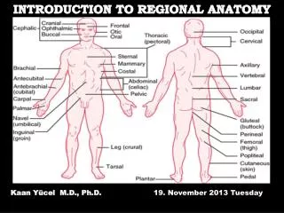
Anatomical Position - supine or prone Anatomical Regions Abdominopelvic quadrants and abdominopelvic regions Superficial Anatomy
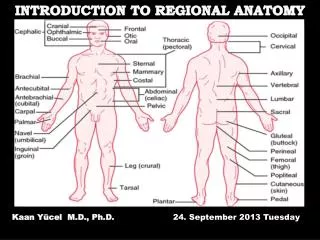
Frons or forehead (frontal) Nasus or nose (nasal) Anatomical Landmarks Oculus or eye (orbital or ocular) Cranium or skull (cranial) Auris or ear (otic) Cephalon or head (cephalic) Facies or face (facial) Bucca or cheek (buccal) Cervicis or neck (cervical) Oris or mouth (oral) Thoracis or thorax, chest (thoracic) Mentis or chin (mental) Axilla or armpit (axillary) Mamma or breast (mammary) Trunk Brachium or arm (brachial) Abdomen (abdominal) Antecubitis or front of elbow (antecubital) Umbilicus or navel (umbilical) LE 1-8a
Antebrachium or forearm (antebrachial) Pelvis (pelvic) Carpus or wrist (carpal) Manus or hand (manual) Palma or palm (palmar) Inguen or groin (inguinal) Digits (phalanges) or fingers (digital or phalangeal) Pollex or thumb Pubis (pubic) Patella or kneecap (patellar) Crus or leg (crural) Femur or thigh (femoral) Tarsus or ankle tarsal) Digits (phalanges) or toes (digital or phalangeal) Pes or foot (pedal) Hallux or great toe LE 1-8a
Cephalon or head (cephalic) Shoulder (acromial) LE 1-8b_1 Cervicis or neck (cervical) Dorsum or back (dorsal) Olecranon or back of elbow (olecranal) Upper limb Lumbus or loin (lumbar)
LE 1-8b_2 Gluteus or buttock (gluteal) Popliteus or back of knee (popliteal) Lower limb Sura or calf (sural) Calcaneus or heel of foot (calcaneal) Planta or sole of foot (plantar)
To facilitate its study, the abdominopelvic region can be divided into different regions and quadrants There are four quadrants and nine regions - provides clinicians a useful frame of reference Abdominopelvic Region
Abdominopelvic Quadrants Right lobe of liver, gallbladder, right kidney, portions of stomach, small and large intestine Left lobe of liver, stomach, pancreas, left kidney, spleen, portions of large intestine Most of small intestine, and portions of large intestine, left ureter, and reproductive organs (left ovary in female and left spermatic cord in male) Cecum, appendix, and portions of small intestine, reproductive organs (right ovary in female and right spermatic cord in male), and right ureter
Abdominopelvic Regions Epigastric region Left hypochondriac region Right hypochondriac region Umbilical region Right lumbar region Left lumbar region Hypogastric region Right inguinal region Left inguinal region LE 1-9b
R. Hypochondriac - right, upper 1/3; gallbladder, liver, r. kidney Epigastric - Upper, central 1/3; liver, stomach, pancreas, duodenum L. Hypochondriac - left, upper 1/3; spleen, colon, liver, l. kidney, small intestine R. Lumbar - right, lateral 1/3; cecum, ascending colon, liver, r. kidney, small intestine Umbilical - center; umbilicus (navel) is located here; jejunum, ileum, duodenum, colon, kidneys, major abdominal vessels L. Lumbar - left, lateral 1/3; descending colon, l. kidney, small intestine R. Iliac (inguinal) - right, lower 1/3; appendix, cecum, small intestine Hypogastric (pubic) - lower, center 1/3; urinary bladder, small intestine, sigmoid colon, female reproductive organs L. Iliac (inguinal) - left, lower 1/3; small intestine, descending colon, sigmoid colon
Stomach Liver Spleen Gallbladder Large intestine Small intestine Appendix Urinary bladder Superficial anatomical landmarks and underlying organs LE 1-9c
Cranial Right Left Proximal Posterior or dorsal Anterior or ventral Lateral Medial Proximal Caudal Distal Distal Directional Terms LE 1-10
Planes – any slice through a 3 dimensional object can be described through 3 sectional planes - sagittal, transverse, frontal (coronal) Serial reconstruction – a series of sections at small intervals in one sectional plane Body cavities – vital organs are suspended in these internal chambers to provide protection Visible Human Project http://www.nlm.nih.gov/research/visible/visible_human.html Sectional Anatomy
Planes of Section Sagittal plane Frontal plane Transverse plane LE 1-11
Cranial Cavity Vertebral Cavity Superior mediastinum Pleural Cavity Thoracic Cavity Mediastinum with pericardial cavity Diaphargm Ventral Body Cavity Abdominal Cavity Pelvic Cavity (b) Anterior view Body Cavities Dorsal Body Cavity • Cranial Cavity – encases the brain • Vertebral Cavity – runs through the vertebral column encloses the SC Ventral Body Cavity or coelom – contains the viscera 2 major divisions: • Thoracic Cavity - enclosed by the chest wall • Abdominopelvic Cavity - enclosed by the abdominal wall and pelvis
Provides protection; allows organ movement; lining prevents friction LE 1-14 separated by diaphragm into Surrounded by chest wall and diaphragm Contains the peritoneal cavity subdivided into Includes the Contains many digestive glands and organs Contains urinary bladder, reproductive organs, last portion of digestive tract Surrounds right lung Contains the trachea, esophagus, and major vessels Surrounds left lung also contains Surrounds heart
Serous cavity – narrow fluid-filled space lined by a serous membrane (serosa) - Pleura - Pericardium - Peritoneum Parietal serosa – pleura covers the opposing mediastinal surface and inner body wall; pericardium is continuous with the viseral serosa; peritoneum lines the body wall Visceral serosa – covers the visceral organs Serous Fluid – watery lubricant secreted by both serous membranes to minimize friction between organs and cavity walls Mesenteries – organs (stomach, small intestine, parts of large intestine) are suspended by double sheets of peritoneum to provide support and stability with limited movement Serous Cavities
Serous Cavities Visceral pericardium Heart Pleural cavity Pericardial cavity Thoracic cavity Pleural cavity Parietal pericardium Pericardial cavity Pericardial cavity Diaphragm Air space Diaphragm Balloon Abdominal cavity Peritoneal cavity Abdominal cavity Abdominopelvic cavity Pelvic cavity Pelvic cavity Sternum Heart in pericardial cavity Pleural cavity Right lung Left lung Left lung Right lung Pleura Mediastinum LE 1-13 Spinal cord
CT, CAT (computerized [axial] tomography) Diagnosis and prognosis Disease MRI (magnetic resonance imaging) Pathology Radiologist Ultrasound X-rays and radiodensity (air, fat, liver, blood, muscle, bone) Clinical Terms
Stomach Small intestine X-Ray Barium-contrast X-ray LE 1-15a
Stomach Liver Aorta Spleen Left kidney Right kidney Vertebra Stomach Liver Aorta Stomach Rib Liver Stomach Liver Left kidney Kidney Vertebra Kidney Kidney Spleen Vertebra Spleen Ultrasound - echogram CT Scan LE 1-16 MRI
Digital Subtraction Angiography (DSA) Spiral CT Sternum Heart First rib Aortic arch Heart Arteries of the heart Right scapula Vertebral column Aorta LE 1-17