Download
1 / 23
240 likes | 494 Vues
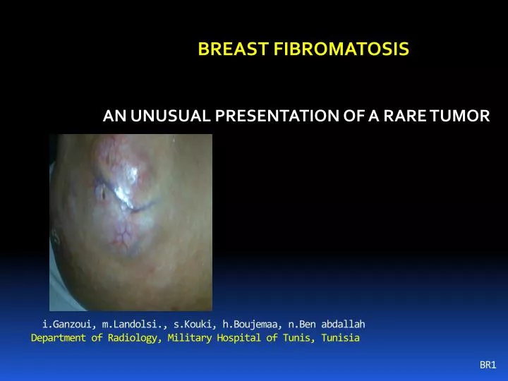
i.Ganzoui, m.Landolsi., s.Kouki, h.Boujemaa, n.Ben abdallah Department of Radiology, Military Hospital of Tunis, Tunisia BR1. BREAST FIBROMATOSIS AN UNUSUAL PRESENTATION OF A RARE TUMOR . INTRODUCTION.

E N D
i.Ganzoui, m.Landolsi., s.Kouki, h.Boujemaa, n.Ben abdallah Department of Radiology, Military Hospital of Tunis, Tunisia BR1 BREAST FIBROMATOSIS AN UNUSUAL PRESENTATION OF A RARE TUMOR
INTRODUCTION Breast fibromatosis (BF) is a rare condition representing less than 0.2% of primary tumors of the breast. It is a benign entity but it is locally aggressive with a recurrence rate reaching 29%.
INTRODUCTION It usually manifests as a painless, unilateral nodule with a hard consistency and irregular contours, with an average size of 3 cm. The diagnosis is based on histological examination which shows a fibroblasts and myofibroblasts proliferation.
Breast Fibromatosis mimics malignancy both clinically and radiologically, it is usually described as an ACR 4 or ACR 5 lesion
OBSERVATION .A 43-year-old woman presented with right breast lumps lasting for one year with no symptoms of impaired general condition. .the entire right breast was the set of confluent large masses measuring approximately 25cm on the great axis, of hard consistency, distorting its shape. .The left breast was with no abnormalities. . both axillae werenormal.
MAMMOGRAPHY .The realization of the right breast mammogram was technically not possible because of its volume. .No focal abnormality was noted in the left breast.
ULRASONOGRAPHY objectified heterogeneous confluent ill- defined formations with posterior acoustic enhancement slightly vascularized on the color- Doppler.( Fig 1 and 2).
MRI 3T Protocole: Due the technical difficulties with such voluminous breast we couldn`t use dedicated breast surface coils. An abdominal phased array antenna was used instead. Axial T1 and T2 weighted images (WI) Sagittal T1 and T2 WI Dynamic 3D T1 Fat Sat injection Post treatment: MIP, Soustraction and enhacement Pattern .
MRI the tumor showed on T1 weighted images a hypo signal similar to the muscle signal. A hyper signal on T2 weighted images A progressive heterogeneous enhancement after contrast material administration. There was no parietal extension or axillary lymph nodes.
Three foci were selected as regions of interesttype I Pattern. progressive enhancement,with a continuous increase in signal intensityon
TREATMENT Due to the tumor volume, a complete mastectomy was necessary. Extemporaneous histological examination showed a benign tumor with healthy limits of the section. The definitive examination of the specimen concluded to mammary fibromatosis The patient remains disease-free with a 5-month follow-up.
DISCUSSION • Most of the reported cases in the litterature were described as a single movable hard or firm mass. • Skin retraction and fixation to the muscle are often present • The sonographic appearance of this disease is typically an irregular and ill-defined hypoechoic mass that is indistinguishable from breast cancer . • although unusual benign-like sonographic findings of well-defined hypoechoic breast nodules have also been reported.
DISCUSSION It is important to recognize mammary fibromatosis. Microbiopsy , although not entirely specific, may be a source of important information . It confidenty allows the exclusion of breast cancer and other more common diseases and is useful in planning a surgical approach to the lesion. None of the literature we reviewed reported such voluminous case of Breast Fibromatosis.
CONCLUSION Breast Fibromatosis is a rare benign entity, mimicking breast cancer . Awareness of such lesions by radiologists, surgeons and pathologists is therefore of paramount importance to guard against an unnecessarily drastic surgical approach, as a complete localized excision is all that is required.
References 1- C. Salem et al. Affections rares du sein. EMC Radiologie et imagerie médicale 2011. 2- R. Al-Yusuf el al. BreastFibromatosis. Bahrain Med Bull2005; 27(4): 1-7. 3- A. Aubertin et al. MammaryFibromatosis: report of five cases and literaturereview. J Le Sein 2004;14:269-278. 4- C. Corbisier et al. Tumeur rare du sein : la fibromatose mammaire. J GynecolObstetBiolReprod 1997;26:315-320. 5- S. Croce et al. BreastFibromatosis: an uncommonbenignbreastdisease. Gynécologie Obstétrique et Fertilité 2009;37:442-446. 6- H. Roman et al. Fibromatose primitive du sein. Ann Chir 2001;126:561-4. 7- M. Driss et al. Fibromateuse mammaire : étude anatomo-clinique à propos de deux observations. Ann Pathol 2002;22:206-9. 8- M. Devouassoux-Shishebouran et al. An infiltrativebreastlesion not to bemisdiagnosed. Ann Pathol 2003;23:615-6. 9- L. Kribi et al. La mastopathir fibreuse diabétique. J Radiol 1999;80:1579-81.