Download

1 / 38
460 likes | 1.56k Vues
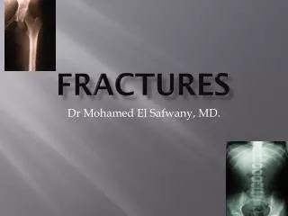
MONTEGGIA AND GALEAZZI FRACTURES. ANATOMY-ELBOW. Hinge joint. Three bones form the elbow joint: the humerus of the upper arm, and the paired radius and ulna of the forearm.

E N D
ANATOMY-ELBOW • Hinge joint. • Three bones form the elbow joint: the humerus of the upper arm, and the paired radius and ulna of the forearm. • The bony prominence at the very tip of the elbow is the olecranon process of the ulna, and the inner aspect of the elbow is called the antecubital fossa.
Humeroulnar joint- **from trochlear notch of the ulna **to trochlea of humerus • Is a simple hinge-joint, and allows of movements of flexion and extension only.
Humeroradial joint- **from head of the radius **to capitulum of the humerus • Is a hinge-joint
Proximal radioulnar joint. **From-head of the radius **to radial notch of the ulna • pronation and supination.
Ligaments:- • Ulnar collateral ligament, • Radial collateral ligament, and • Annular ligament.
The muscles in relation with the joint are: in front, the Brachialis, the Brachioradialis behind, the Triceps brachii and Anconæus laterally, the Supinator, and the common tendon of origin of the Extensor muscles medially,-common tendon of origin of the Flexor muscles, and the Flexor carpi ulnaris
Movements • The hinge-like bending and straightening (flexion and extension) between the humerus and the ulna. • The complex action of turning the forearm over (pronation or supination) happens at the articulation between the radius and the ulna (this movement also occurs at the wrist joint). • The hinge moves in only one plane.
The Arteries supplying the joint are derived from the anastomosis between the profunda and the superior and inferior ulnar collateral branches of the brachial, with the anterior, posterior, and interosseous recurrent branches of the ulnar, and the recurrent branch of the radial. These vessels form a complete anastomotic network around the joint. • The Nerves of the joint are a twig from the ulnar, as it passes between the medial condyle and the olecranon; a filament from the musculocutaneous, and two from the median.
Monteggia fracture • # of upper third of ulna with dislocation of head of radius. • Head of radius is dislocated both from the radioulnar articulation and from elbow joint. • It may be displaced –Ant,post,or laterally acc to angulature of ulnar fracture.
DIAGNOSIS • Every # of upper shaft of ulna without # of radial shaft should be considered to be monteggia # unless otherwise proved. • first X ray may show head of radius in its correct position, but serial X rays have to be taken over 1st few weeks –bcoz if dislocation has occurred and there is instability ,head of radius may redisplace later.
Displacement-3 types • Monteggia # dislocations can take place from 3 forces and corresponding injuries seen. • FLEXION INJURY • EXTENSION INJURY • ADDUCTION INJURY • ***Hume fracture
FLEXION INJURY-10-15% • # ulna is angulated with the convexity posteriorly and the head of radius is dislocated backwards.
EXTENSION INJURY-85-90% • Commonest type. • # ulna is angulated with covexity ant. and laterally. • With head of radius dislocated forwards and laterally.
Adduction injury • Caused by adduction strain at the elbow. • Ulna is angulated laterally and radial head is displaced laterally.
HUME FRACTURE • “High Monteggia injury”. • 1957 Hume described --fracture of the olecranon with an associated anteriordislocation of the radial head . • Seen in Children.
MECHANISM OF INJURY. • Mervyn Evans suggested this mech. • 1**Fall on outstretched hand with twisting of the trunk,forcibly pronating the forearm. • 2**Direct injury-Africa-Direct blow on the back of forearm with a stickwhile arm is raised warding off an attacker.
TREATMENT • CONSERVATIVE • OPERATIVE
CONSERVATIVE:- • Children. • manipulation and plaster immobilisation. • But close watch needed-recurrence of deformity.
Redn. of extension injury. • Longitudinal traction of forearm with with the elbow flexed as much as possible without compromising the blood supply. • Forearm is stable in supination • Plaster windowed for radial pulse
Redn of adduction injury. • Traction of the forearm with elbow extended and pressure over the head of radius, and after redn.this # dislocation is stable with the elbow flexed.and with forearm supinated.
Redn of flexion injury • Traction on forearm with elbow extende and as the redn is stable only in the extended position –not advisable in adults.
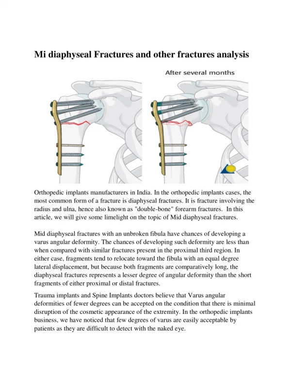
OPERATIVE TREATMENT. • Advisable in adults. • Open redn of # ulna and rigid int. fixation preferable with a plate.. • Dislocation of head of radius red. spontaneously when the deformity of ulna has been reduced.
OPERATIVE TECHNIQUE. • # of ulna is exposed ,reduced and fixed by a compression plate,or IM nail. • Intraop take xray elbow in 2 planes. • If head of radius is perfectly reduced, the position is accepted and well padded plaster cast is applied from metacarpals to axilla- with elbow at right angles and forearm supinated.
If X ray shows –head of radius is not reduced, then it must be exposed and reduced under direct vision. • Annular lig. --usually cause obstruction-incised.
COMPLICATIONS 1.UNREDUCED DISLOCATION OF HEAD OF RADIUS. 2.TRAUMATIC OSSIFICATION AROUND RADIAL HEAD. 3.PIN PALSY 4.CROSS UNION B/W RADIUS AND ULNA. 5.DISLOCATION OF LOWER END OF ULNA 6.UN-UNITED # OF ULNA.
Unred. disl. of head of radius. • Rx • Excision of displaced head of radius. • Prod inc. elbow flexion and good range of pronation and supination. • NOT done in CHILDREN.—removal of upper radial epiphysis—inequality of length of forearm bones and cause further disl. of RU joints both sup. and inf.
Traumatic ossi. around radial head. • Excision of radial head and the block of bone attached to it. • Recurrence. • Can be reduced by Sx delayed 6-12 months after injury with elbow immobilised for atleast 2 weeks. • NO Physiotherapy,manipulation and passive excs during rehab period.
PIN PALSY • Common with Adduction # dislocation. • Prognosis good in early complete reduction of head of radius. • Late PIN palsy due to inadequate redn of radial head.
Cross union b/w radius and ulna. • Bony fusion b/w neck of radius and 3 site of upper 3rd of ulna. • Difficult to Rx. • B coz proximity of elbow jt and PIN. ***Recurrence is high. • ***Perm limitation of Radioulnar movt.
Dislocation of lower end of ulna • REDUCES with redn of ulnar shaft #. • WORSENS if head of radius is excised. • Rx excise distal inch of ulnar-if wrist symptoms.
Un united # of ulna • Notorious for that. • Rigid internal fixation and cancellous onlay grafting.