Download
1 / 27
280 likes | 321 Vues
In this chapter on Cell Growth, Division, and Reproduction, discover why cells must divide, the types of cell division, and the necessity of the cell cycle for growth, repair, and reproduction. Explore the processes of mitosis and meiosis, learn about regulation mechanisms, and understand the implications of uncontrolled cell division in cancer development.

E N D
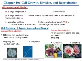
Chapter 10: Cell Growth, Division, and Reproduction Why must a cell divide? a) a large cell places a large demand on the DNA… “info overload” b) a large cell has a low surface area to volume ratio – cell is less efficient at moving materials in and out c) a smaller cell has it’s own DNA – (not overworked anymore) AND a HIGH surface area to volume ratio. Can manage cell needs better Cell Division: 2 Types: Asexual and Sexual • Sexual Reproduction: • Fertilization of sperm and egg produces genetically different offspring • Creates a variety of offspring Asexual Reproduction: * Offspring are produced by a single parent, without the participation of sperm and egg * offspring are genetic copies of the parent and of each other
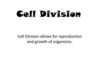
Why is cell division necessary? growth cell replacement wound repair asexual reproduction
Prokaryotic chromosomes Colorized TEM 32,500 Prokaryotes reproduce by binary fission: asexual • As the cell replicates its single chromosome, the copies move apart • The growing membrane then divides the cells Figure 8.3B
LM 600 THE EUKARYOTIC CELL CYCLE AND MITOSIS The large, complex chromosomes of eukaryotes duplicate with each cell division • A eukaryote has many more genes (and chromosomes) than a prokaryote • Genes are grouped into multiple chromosomes in the nucleus • Each chromosome contains a very long DNA chain with attached histone proteins • Chromosomes are ONLY visible during cell division • If a cell is not undergoing division, chromosomes unwind into loosely packed fibers called chromatin 46 chromosomes unwound = 2 meters!
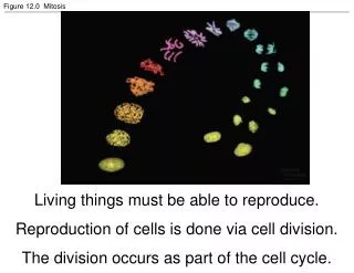
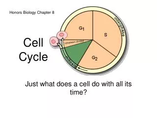
The Cell Cycle: Interphase and Mitosis • 1. Interphase:All DNA is duplicated and cell parts are made. 3 stages: • G (gap)1: normal cell growth • S (synthesis): all DNA is copied (new set of chromatin is synthesized) • G (gap)2: organelles replicated • Mitosis:Duplicated • chromosomes are evenly • distributed into two daughter • nuclei. 4 phases: • Prophase • Metaphase • Anaphase • Telophase Cytokinesis
The Stages of Mitosis: = centromere Sister chromatids Interphase: G1, S, and G2 Prophase: chromatin condenses, sister chromatids join, nucleolus disappears, centrosomes (centrioles) start to produce spindle fibers and begin migrating to poles, nuclear envelope breaks down, spindles attach to chromosomes, and start moving them to the middle
The Stages of Mitosis: cont’d Mitosis video Metaphase: chromosome pairs and centromeres are lined up at middle Anaphase: centromeres split, spindles which are attached to chromosomes recoil and split sister chromatids apart, other spindles get longer and poles are pulled farther apart…cell is stretched Telophase and Cytokinesis: chromosomes unwind into chromatin, nuclear envelope and nucleolus form, spindles disappear, microfilaments pinch at center cytokinesis…cytoplasm completely divides (in plants a cell plate is formed)... 2 new cells
SUMMARY: Putting it all together Drawings ANDReal Pictures
Cytokinesis differs for plant and animal cells • In animals, cytokinesis occurs by a constriction of the cell (cleavage) • In plants, cell membrane can’t pinch b/c of cell wall. A cell plate forms instead Figure 8.7A
Do all cells divide at the same rate?? NO Fast dividing: intestinal lining, bone marrow, skin, follicle cells Slow dividing: liver, pancreas Non-dividing: nerve and muscle (after about 5 years of age) Q: How is the cell cycle regulated in each type of cell? A: Physical boundaries, Cyclins, and Regulatory Proteins 1. Physical Boundaries Most cells stop dividing once they TOUCH each other ex) cells in a petri dish grow in a single layer and stop once whole surface is covered. Remove some cells, and border cells will divide again to fillthe gap. ex) woundboundary cells are stimulated to divide until the space (wound) is healed.
Regulation of Cell Cycle: Cyclins and Regulatory Proteins 2. Cyclins: proteins present in cells ONLY when dividing 3. Regulatory Proteins: used IN and OUT of the cell to stimulate division ex) INternal regulatory proteins: * one type makes sure all chromosomes are made before going further * another type makes sure all spindles are formed before going further ex) EXternal regulatory proteins: direct cells to increase OR decrease RATE of cell division * growth factors – outside chemical signal for cell to start division: vital in embryo developmentand wound healing * others work as “red lights” to slow down cell division – preventing tumor development
Cancer: Uncontrolled Cell Division Cancer cells divide excessively; no control system • produce malignant tumors which invade normal cell space, robbing them of nutrients and blood supply • produce own growth factors constant divide signal • divide and live longer than normal cells • Tumor: benign (cells stay put) OR malignant (cells metastasize) • Carcinomas (covering and linings): skin, intestine (colon) • Sarcomas (support tissue): muscle, bone • Leukemia and Lymphoma: blood tissue • Chemotherapy: side effects felt most by fast-dividing cells • ex: hair follicles, intestinal lining, immune cells • Anti-Cancer drugs: all botanical extracts * Taxol: freezes mitotic spindle no division * Vinblastin and Colchicine: stop spindle formation no division
Q: What causes cancer cell growth ? A: defects (mutations) in the DNA (genes) which code for regulatory proteins How do these defects happen?? 1. chemical exposure (ex: tobacco and cigarette smoke) 2. radiation exposure (ex: x-rays, uv light rays from sun) 3. viruses can disrupt DNA (ex: HPV, HIV) ** Many cancer cells have a mutation in gene “P53” “P53” gene codes for an internal regulatory cell cycle protein at the S of Interphase.
Normal, Programmed, Cell Death?? Apoptosis: pre-programmed, deliberate cell death Process: 1. cell looses fluid and shrinks 2. chromatin breaks down 3. cell membrane distintegrates 4. cell’s neighbors clean up debris and re-use materials Why needed? 1. This is a key role in embryonic development (ie; shape changes) ex: paddle hand ex: tadpole frog ** Too much cell death is linked to certain diseases (Parkinson’s, AIDS)
CHROMOSOMES Chromosomes: coiled DNA (chromatin) • in humans: 23 pairs 46 in ea. body cell (23 from mom, 23 from dad) • Homologous chromosomes: same #, size, shape, gene location, centromere location (ex: chromosome #1 from mom is homologous to chromosome #1 from dad, etc.) • haploid # = (n) half (23 total): sex cells (egg and sperm) • diploid # = (2n) double (46 total): body (somatic) cells • Sex cells (23, haploid): 22 autosomes (#1 #22), 1 sex chromosome (#23) • Body cells (46, diploid): 22 pairs of autosomes… (44 total) 1 pair of sex chromosomes (XX) or (XY) • Q: Which cell determines the sex of the offspring; egg or sperm?
MEIOSIS: a.k.a. “reduction division” • Summary: • Diploid (2n) cell(germ cell) forms cells with (n) nuclei • Two divisions • Used for gamete • formation only • Takes much longer • than mitosis • Phases: • Interphase (G1, S, G2) • Prophase I • Metaphase I • Anaphase I • Telophase I & Cytokinesis • Prophase II • Metaphase II • Anaphase II • Telophase II • & Cytokinesis
Stages of Meiosis Unique features animation • Interphase: G1, S, G2 • Prophase I: • chromatin condenses • sister chromatids join at centromere • homologous pairs of sister chromatids join to make tetrads • crossing over occurs to shuffle genes • nuclear envelope and nucleolus disappear • centrioles migrate, grow spindles, attach to tetrads • tetrads start to move to middle of cell • Metaphase I: tetrads lined up at metaphase plate • Anaphase I: • spindles recoil • homologous pairs pulled apart • each pair of sister chromatids move to opposite pole. • Telophase I and Cytokinesis: • cell pinches in middle divide cytoplasm. • each cell (2) has 1 pair of sister chromatids fromeach original tetrad
Stages of Meiosis cont’d • Prophase II: • spindles form again, • spindles attach to and start moving sister chromatid pairs to middle • Metaphase II: • sister chromatid pairs lined up at • metaphase plate • Anaphase II: • spindles recoil • centromeres divide • sister chromatids separate • Telophase II and Cytokinesis: • nucleolus and nuclear envelope appear • chromosomes unwind into chromatin • cell pinches in middle • divide cytoplasm…4 new daughter cells • each with (n) number Meiosis stages video
comparison animation Review: A comparison of mitosis and meiosis
Gamete formation begins with a 2n body cell Oogenesis: production of egg (ovum) Oogonia: female germ cells (2n) which will undergo meiosis and make 1 egg (n) and 3 polar bodies (n) each time Spermatogenesis: production of sperm (spermatozoa) Spermatogonia: male germ cells (2n) which will undergo meiosis to make 4sperm (n) cells each time s s s s EGG 3 polar bodies Polar bodies: a waste of DNA in order to get a large egg • Spermatozoa: small, very little cytoplasm, flagella • Head with nucleus, midpiece with mitochondria, flagella conservation of cytoplasm:1 big egg with lots of energy in it’s stored food
Q: Why is everyone different? (except identical twins) • Random crossing over of genes during prophase I • Random orientation of chromosomes at metaphase plate • Random fertilization All this leads to many different combinations of chromosomes in eggs and sperm Orientation animation 2 23 = 8 mill combo’s for egg/sperm at fertilization: 8M x 8M = 64 trillion combo’s of chromosomes…not including crossing over!!
Alterations of chromosome number and structure Karyotype: a photographic inventory of an individual’s chromosomes • used to detect abnormal chromosome number or abnormal chromosome shape non-disjunction: failure of chromosomes to separate equally • Can lead to trisomy (2n + 1) individual • Trisomy 21 : Down’s Syndrome ( 1 in 700 births; most common) • Trisomy 18, 15, etc… • XYY: Super Male • XXX: Super female • XXY : Klienfelter’s Syndrome • XO: Turner’s Syndrome Trisomy 21
Non-disjunction in Meiosis I vs. Meiosis II 0 • failure of homologous pairs to separate • failure of sister chromatids to separate
Polyploidy: Extra Set(s) of Chromosomes • Results when entire set (s) of chromosomes fail to separate (nondisjunction) during meiosis • 2n + n = 3n (Triploid) • 2n + 2n = 4n (Tetraploid) • Polyploidy is lethal in humans but is tolerated in plants • Plants can be forced into polyploidy with colchicine • ex. polyploid coffee beans provide more robust flavor Miscarriage, severe birth defects, or early death
Alterations of chromosome structure: birth defects and cancer • Deletions, duplications, inversions, and translocations • Occurs during crossing over example
Bonus Questions: Pick any 2 of your choice • What is the full name of a sperm cell? • During which phase is non-disjunction of chromosomes most problematic (Meiosis I or Meiosis II)? • When a karyotype is made, what is the “n” number of that cell (n, 2n, 3n or 4n)? • Name a chemotherapy agent which prohibits spindle formation. • What term describes the spread of malignant cancer to other organs in the body? • Cancer of the tissues which function as coverings and linings are collectively called?