Download
1 / 8
90 likes | 261 Vues
IBIC Summer Retreat Allen Institute Human Brain Atlas. Allan R. Jones, PhD Chief Scientific Officer June 7, 2009. Allen Institute for Brain Science: Fueling Discovery. Who/what we are:

E N D
IBIC Summer RetreatAllen Institute Human Brain Atlas Allan R. Jones, PhD Chief Scientific Officer June 7, 2009
Allen Institute for Brain Science: Fueling Discovery • Who/what we are: • An independent, non-profit research organization working to support basic research in the brain sciences (founded in 2001). • Dedicated to making tools and information readily available to the scientific community • Project-focused, milestone driven • Multi-disciplinary teams working towards a common goal (math, physics, engineering, systems-level and molecular neuroscience, molecular biology, genetics, genomics, information technology) • ~120 staff (30 PhDs) • Located in 35000 sf of mixed lab/office space in the Fremont neighborhood of Seattle Washington • What we are not: • A traditional, PI-driven research organization • An extramural funding agency
The Allen Institute for Brain Science: Tools, resources, and data • Atlases: • Adult mouse brain (complete Sept. 2006) • Mouse spinal cord (complete April 2009) • Mouse development (complete March 2010) • Human brain • Phase 1 complete mid-2010 • Phase 2 complete in 2012 • Projects: • Sleep study (complete Dec. 2007) • Genetic diversity study (complete May 2008) • Human cortex survey (complete Sept. 2008) • Human cortex population study/schizophrenia (complete Jan. 2009) • Human Glioblastoma (complete April 2011 with possible extension) • Tools: • Transgenic mouse drivers/reporters • ~20,000 unique visits per month across all projects (65% mouse brain, 10% human cortex, 10% development, 10% spinal cord, 5% sleep) • Atlas paper has been cited 260 times since publication in January 2007 (validation, discovery)
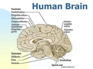
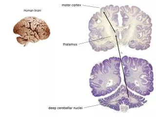
Connecting the “what” to the “where” • We are quickly approaching a renaissance in our understanding of the basic genetic underpinnings of human biology and behavior • Technology has enabled easy, cheap access to high resolution genetic data from humans. Large scale studies are underway • Technology has provided ways to link functions in the brain to location • Researchers that study genetics of human behavior and brain disease will be able to identify key genes • Researchers that study brain function can already pinpoint brain locations that are altered or perform aberrantly in disease • A key resource is needed to tie the “functional” maps with the “genetic” maps: a gene expression map of the human brain
2008 2009 2010 2011 2012 Initial data release Project completion Status Check Protocol v 0.5 Planning Phase 1: Microarray Data Planning Phase 2: ISH data Human Brain Atlas Overview: 2008-2012Multimodal atlas integrating gene expression and neuroanatomy • Phase 1: Anatomic resolution atlas • All structures: • Comprehensively sample human brain • All genes: • Microarray and sequence-based gene expression profiling • Phase 2: Cellular resolution atlas • Most structures: • High-resolution atlas of each structure • High-value genes: • ISH for 50-500 genes/structure
5 mm coronal slabs Section 1 mm of slab Full coronal histology Whole brain MRI Myelin Myelin Myelin Myelin Myelin Myelin Nissl Nissl Nissl Nissl IHC IHC IHC LCM ISH IHC IHC IHC IHC IHC IHC IHC Nissl Nissl Whole brain to microarray: serial divisions (Fresh brain images courtesy Mark Vawter and Preston Cartagena) Cryosection through 3 mm of 2x3 block Final 1 mm of block Subdivide slab into 2x3 blocks cortex 1 cm sampling gross dissection subcortex 0.5 cm sampling (subset of ~60 structures) ~700 cortical samples ~300 subcortical samples
MRI and DTI 2-10 human brain specimens Anatomic segmentations (a) (b) (c) Macro-dissection (cortex) Microarray analysis Subdivided slab 2x3 blocks 5 mm coronal slabs LCM (sub-cortex) (d) (i) (g) (h) 6x8 histology, 2x3 histology, immunohistochemistry and in situ hybridization (ISH) User applications Blockface images (f) (j) (e)