Saladin Ch. 22
1.32k likes | 1.59k Vues
Saladin Ch. 22. Respiratory System. Alveoli. Respiratory Processes. Pulmonary ventilation - Inspiration and expiration External respiration - O 2 into pulmonary circulation, CO 2 out. Transport of gases. Respiratory Processes.

Saladin Ch. 22
E N D
Presentation Transcript
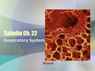
Saladin Ch. 22 Respiratory System Alveoli
Respiratory Processes • Pulmonary ventilation - Inspiration and expiration • External respiration - O2 into pulmonary circulation, CO2 out. • Transport of gases
Respiratory Processes • Internal respiration - O2 out of capillaries, CO2 in. • Cellular respiration - metabolic reactions within cells that consume O2 and produce CO2.
Functions • Gas exchange - O2 in, CO2 out • Regulation of blood pH • Sense of smell • Filtering system for inspired air
Functions • Sound production for communication • Heat and water reduction
Respiratory System Components • Upper Respiratory system = nose, nasal cavity, pharynx and associated structures. • Lower Respiratory System = larynx, trachea, bronchi and lungs.
Respiratory System Components • Conducting - nose, pharynx, larynx, trachea, bronchi, bronchioles, & terminal bronchioles - filter, warm & moisten air and carry to the respiratory portion. • Respiratory Portion - tissues within lungs where gas exchange transpires.
Nose • Functions - warm, moisten, filter air, smell, modifying speech sounds. • External – Nose • Structures – root, bridge, dorsum nasi, apex, philtrum. • Bones - nasal, frontal, and maxilla.
Nose • Cartilage - septal, lateral nasal and alar. • External nares - openings into external nasal region from outside.
Nose • Nasal cavity - space inside internal nose. Divided into R and L by nasal septum. • Anterior portion is the vestibule - has nasal hairs.
Nose • Internal – nasal cavity • Bones - ethmoid, maxilla, lacrimal, palatines, nasal conchae. • Internal nares - openings from internal nasal region into pharynx. • Ducts from paranasal sinuses, & nasolacrimal glands enter here.
Nose • Superior, middle & inferior meatuses. • Formed by coverings over the nasal conchae. • Mucous membranes - olfactory epithelium in superior.
Nose • Erectile tissue – inferior concha – swells and closes nostrils – alternates a couple of time per hour to allow for recovery from drying. • Pseudostratified columnar epithelium with goblet cells - sweep and weep.
Pharynx • Throat - from internal nares to cricoid cartilage [most inferior of laryngeal cartilages]. • Skeletal muscle wall lined with mucus membranes. 3 regions.
Pharynx • Nasopharynx- from nasal cavity to plane of soft palate. • Pseudostratified columnar epithelium. • 5 openings - 2 internal nares, 2 auditory (Eustachian tubes), opening into oropharynx.
Pharynx • Receives air from nares & mucus packets of trash to be removed. • Also helps equalize ear pressure via the Eustachian tubes. • Pharyngeal tonsils [adenoids] trap & destroy pathogens.
Pharynx • Oropharynx - from nasopharynx to epiglottis. • Common passageway for food, drink & air - digestive & respiratory. • Stratified squamous epithelium - to withstand food abrasion. • Lingual & palatine tonsils.
Pharynx • Laryngopharynx - connects esophagus to voice box. • Stratified squamous epithelium. • Epiglottis to larynx.
Larynx • Connects pharynx to trachea - C4-C6 region. • From top to Bottom: • Hyoid bone • Thyrohyoid membrane
Larynx • Thyroid cartilage • Behind this: • Cuneiform (2) • Corniculate(2) • Arytenoid cartilage (2)
Larynx • Cricothyroid ligament • Cricoid cartilage • Cricotracheal ligament
Larynx • Epiglottis/glottis: • Epiglottis attached to thyroid cartilage. • Covers glottis (vocal cords & opening between) during swallowing - to prevent stuff from going the wrong way.
Larynx • Linings: • Above larynx = non-keratinized stratified squamous. • Below -pseudostratified columnar with cilia and goblet cells.
Structures of Voice Production • Vestibular folds [false vocal cords - superior] - hold breath in thoracic cavity by bringing folds together.
Structures of Voice Production • Vocal folds [true vocal cords] • Bands of elastic ligaments stretched between pieces of cartilage - like guitar strings. • Skeletal muscles move the vocal folds into & out of the air stream & tighten or loosen the "strings“. • More air - more volume.
Structures of Voice Production • Shorter strings produce higher pitch. • Men tend to have longer, thicker "strings" thus lower voices. • Shape of the resonating chamber affects intonation, etc. - like with any instrument. • Cheeks, tongue, lips etc. contribute.
Trachea • From larynx to fifth thoracic vertebra. • 12 cm long, 2.3 cm diameter. • Anterior to esophagus. • Passageway for air and filters air.
Trachea • Layers: • Mucosa [pseudostratified ciliated columnar epithelium with goblet cells. • Submucosa - contains ducts and glands; connective tissue & muscle between ends = trachealis muscles.
Trachea • Adventitia - outer layer - loose connective tissue. • Cartilagenous layer - 16 to 20 c-shaped rings with transverse smooth.
Trachea • Tracheostomy - incision into trachea just inferior to cricoid cartilage - then place a tube. • Intubation - the ramrod approach - just shove any obstruction down the tube, then suction out the mess.
Bronchi • Trachea branches into R & L primary bronchi. • Carina - branch point - very sensitive - has cough reflex. • R is more vertical, shorter & wider - more likely to get inhaled objects. • L is longer and narrower.
Lungs • External Anatomy Features: • Base, apex, hilus [where vessels, bronchi, etc. enter each lung]. • Costal surfaces, cardiac notch.
Lungs • Fissures - divide the lungs into lobes. • Oblique - in both divides into superior and inferior lobe. • Horizontal - R only; splits the superior to form a third, medial lobe.
Bronchi • Anatomy: • Surrounded by incomplete rings of cartilage like trachea. • Lined with pseudostratified ciliated columnar epithelium.
Bronchi • Branching: • Primary bronchus secondary bronchi - to each lobe - 3 to R & 2 to L. • Secondary tertiary – to 10 [R] or 8 [L] bronchopulmonary segments per lung.
Bronchi • Tertiary branch to bronchioles that branch to terminal bronchioles, etc. • Total of 25 branching divisions. • Pulmonary arteries & branches parallel bronchial tree