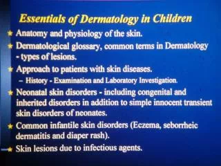
Milia
Primary lesions are de-novo lesions while secondary are either a sequence of the natural history of the disease or a modification of the primary lesion which can be either due to drug use or iatrogenic by itching.

Milia
E N D
Presentation Transcript
Primary lesions are de-novo lesions while secondary are either a sequence of the natural history of the disease or a modification of the primary lesion which can be either due to drug use or iatrogenic by itching. • Dermatological test of choice is skin biopsy and it is done whenever we are in doubt.
Milia • Its an epidermal inclusion cyst. • Simple benign and asymptomatic. • Unknown cause. • Seen as papules, in the next picture they are over the cheeks and nose. • Papules contain keratinized material. • Resolves spontaneously within days.
Ebsteinpearls It is an epidermal inclusion cysts, it affects mucous membrane and most commonly the mouth. When it ruptures it involutes spontaneously.
Ranula It is a congenital mucous retention thin cyst, less commonly traumatic. It ruptures spontaneously and needs no surgical intervention.
Erythema Toxicum Neonatorum • It is a macular generalized patchy red rash. • Benign and self limiting. • Ruptures and resolves spontaneously. Therefore reassure the patient. • Patient does not look sick and has no fever. • Macules are the commonest presentation, but can also present with papules and vesicles. • It is an esinophilic eruptions. • Scraping taken from the lesion will show esinophils aggregate. • The next picture also shows infantile gynaecomastia, which is physiological due to the maternal hormones. It can also produce milk. It resolves spontaneously when the estrogen levels go down in the blood. Do not squeeze it or touch it because you might induce mastitis.
Congenital Macular rash that appears on the back of the neck, glabella and the upper eyelid. Its an ictatic salmon like rash. It disappears with time especially if above the clavicle or on the upper eyelid. It might also persists for years and then get covered by hair.
Mongolian spots • It occurs anywhere but mostly in the lower back or buttocks. • It is a bluish- greenish discoloration that are caused by the arrest of melanocyte in the dermis. • It is seen in Mongolian race, but it has nothing to do with down syndrome. • Might disappear with time. • Common in our society and dark-skinned people, but less common in Caucasians.
Hemangiomas • It is an abnormal proliferation of the blood vessels. • It might involve the viscera or the respiratory tract. • It appears as papules and increase in size. • Two types: strawberry and capillary hemangiomas. • Strawberry hemangioma: rapidly enlarge within the first two years, and disappears around school age. Therefore it only need reassurance of the patient. It is more benign than the capillary form. • When the strawberry hemangioma enlarges, it can cause bleeding, disfigurement or ulceration. When complications occur, you treat with phototherapy, laser systemic steroids or interferon. • Capillary hemangioma: it most commonly affect the area supplied by the ophthalmic division of the trigeminal nerve. It is usually associated with other symptoms. It never disappear by itself might lead to glucoma (one eye is bigger than the other).
Sturge-Weber syndrome: leptomeningioma with contra-lateral hemiplegia, mental retardation and capillary hemangioma.
KabelKanani syndrome they have hemi-hypertrophy on the side of the hemangioma.
EpidermolysisBullosa: whenever the skin is touched it sloughs out. There is no specific treatment, you need to only prevent infection, and when infection occurs treat it.
Collodion baby AKA Icthyosis Congenita: the skin is very tight the baby can not move or breath. It is a form of icthyosis. The skin will get shed with time but he will continue to have icthyosis, requires supportive manegemnt.
Nevi • They are abnormal cells in an abnormal locations. • Black hairy nevus is AKA giant or trunk nevus. It might covers the whole trunk. The larger it is the more it is likely to convert into malignant melanoma (it increases the risk 10x normal). • All the previous images are congenital skin lesions.
Infantile Eczema- Atopic Dermatitis • Its not present at birth. • It is an inflammatory immunological disorder. There is activation of the mast cells (increased IgE) causing the release of histamine that attacks the skin. • Unknown cause. • It waxes and wanes (intermittent), it might stay for several years. • Usually seen on the flexures. • it causes itching, which will result in secondary lesions as: liquenifacation, ulceration and excoriation. • It requires intermittent long term treatment. • Most common complication is by secondary bacterial infection by staph or strept because the skin barrier is broken. It can also be complicated by eczema herpiticus where herpes virus invade abnormal skin.
Seborrheic Dermatitis • Scaly eythematous oily skin lesions. Has nothing to do with seborrheic glands. • More common in extreme ages. • Involves skin creases as the axilla, face and scalp (cradle scalp-hallmark). • More benign, more superficial and less itchy than atopic dermatitis. • Might disappear and will not reappear until later in life (60-70) years. • Treated by removing the scales, wash properly with shampoo, we might sometimes give mild steroids or salicylic acids or keratolytic drugs.
Contact Dermatitis • Its irritant dermatitis AKA diaper dermatitis. • It spares creases. • Its due to irritation from urine, stool and hot humid environment (as in the diaper). • It might be complicated with fungal infection. In this case the creases will be involved and you might see vesicles. Whenever suspected look and the mouth for oral thrush.