
Microautoradiography Techniques: Analyzing [3H]thymidine Incorporation in Cell Cultures
This document outlines the methodologies for preparing microautoradiographs from cell cultures, emphasizing the incorporation of [3H]thymidine in normal glial cells incubated with a specified concentration. It illustrates examples of typical microautoradiographs, showcasing densely labeled cell nuclei and cytoplasmic labeling in mycoplasma-infected cultures. Additionally, the text touches on somatic cell hybridization and hybridoma production for monoclonal antibody generation, providing insights into cellular interactions and applications in research.

Microautoradiography Techniques: Analyzing [3H]thymidine Incorporation in Cell Cultures
E N D
Presentation Transcript
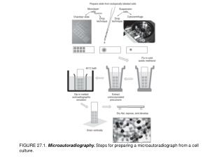
FIGURE 27.1. Microautoradiography. Steps for preparing a microautoradiograph from a cell culture.
FIGURE 27.2. Microautoradiographs. Examples of [3H]thymidine incorporation into a cell onolayer. Normal glial cells were incubated with 0.1 μCi/mL (3.7 KBq/mL), 200 Ci/mmol (7.4 GBq/μmol), of [3H]thymidine for 24 h, washed, and processed as described in Protocol 27.3. (a) Typical densely labeled nuclei, suitable for determining the labeling index (see Protocol 20.11). (b) A similar culture, infected with mycoplasma showing [3H]thymidine incorporation in the cytoplasm. (See also Plates 13c, d, 14d).
FIGURE 27.3. Somatic Cell Hybridization. Selection of hybrid cells after fusion (see Section 27.7).
FIGURE 27.4. Production of Hybridomas. Schematic diagram of the production of hybridoma clones capable of secreting monoclonal antibodies.