PULMONARY EMBOLISM
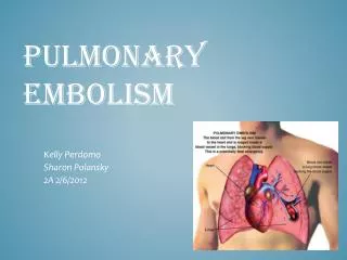
PULMONARY EMBOLISM. Dr. Abdul- Monim Batiha , RN, MSN, PhD. Pulmonary embolism (PE). Pulmonary embolism (PE) refers to the obstruction of the pulmonary artery or one of its branches by a thrombus (or thrombi) that originates somewhere in the venous system or in the right side of the heart.

PULMONARY EMBOLISM
E N D
Presentation Transcript
PULMONARY EMBOLISM Dr. Abdul-MonimBatiha, RN, MSN, PhD
Pulmonary embolism (PE) • Pulmonary embolism (PE) refers to the obstruction of the pulmonary artery or one of its branches by a thrombus (or thrombi) that originates somewhere in the venous system or in the right side of the heart. • Most commonly, PE is due to a blood clot or thrombus.
However, there are other types of emboli: air, fat, amniotic fluid, and septic (from bacterial invasion of the thrombus). • It is estimated that more than half a million people develop PE yearly, resulting in more than 50,000 deaths.
PE is a common disorder and often is associated with trauma, surgery (orthopedic, major abdominal, pelvic, gynecologic), pregnancy, heart failure, age older than 50 years, hypercoagulable states, and prolonged immobility. It also may occur in an apparently healthy person
Risk Factors for Thromboembolism Hereditary Thrombophilias ■ Protein C deficiency ■ Protein S deficiency ■ Antithrombin III deficiency ■ Factor V Leiden mutation ■ Prothrombin 20210 G–A variation ■ Hyperhomocysteinemia Acquired Surgical Predisposition ■ Major thoracic or abdominal surgery requiring general anesthesia and lasting >30 minutes ■ Hip arthroplasty ■ Knee arthroplasty ■ Knee arthroscopy ■ Hip fracture ■ Major trauma ■ Open prostatectomy ■ Spinal cord injury ■ Neurosurgical procedures Acquired Medical Predisposition ■ Prior venous thromboembolism ■ Age >40 years ■ Malignant neoplasia ■ Congestive heart failure ■ Cerebrovascular accident ■ Nephrotic syndrome ■ Estrogen therapy ■ Pregnancy and the postpartum period ■ Obesity ■ Prolonged immobilization ■ Antiphospholipid antibody syndrome ■ Lupus anticoagulant ■ Inflammatory bowel disease
Pathophysiology • When a thrombus completely or partially obstructs a pulmonary artery or its branches, the alveolar dead space is increased. • The area, although continuing to be ventilated, receives little or no blood flow. • Thus, gas exchange is impaired or absent in this area. • In addition, various substances are released from the clot and surrounding area, causing regional blood vessels and bronchioles to constrict.
Pathophysiology • This causes an increase in pulmonary vascular resistance. • This reaction compounds the ventilation–perfusion imbalance.
Pathophysiology • The hemodynamic consequences are increased pulmonary vascular resistance from the regional vasoconstriction and reduced size of the pulmonary vascular bed. • In patients with no preexisting cardiopulmonary disease, obstruction of less than 20% of the pulmonary vascular bed produces compensatory events that minimize adverse hemodynamic consequences.
Pathophysiology • When the degree of pulmonary vascular obstruction exceeds 30% to 40%, increases in pulmonary artery pressure occur, followed by modest increases in right atrial pressure. • When the work requirements of the right ventricle exceed its capacity (the degree of pulmonary artery obstruction exceeds 50% to 60% )right ventricular failure occurs, leading to a decrease in cardiac output followed by a decrease in systemic blood pressure and the development of shock.
Signs and Symptoms of Pulmonary Embolism Small to Moderate Embolus ■ Dyspnea ■ Tachypnea ■ Tachycardia ■ Chest pain ■ Mild fever ■ Hypoxemia ■ Apprehension ■ Cough ■ Diaphoresis ■ Decreased breath sounds over affected area ■ Rales ■ Wheezing Massive Embolus A more pronounced manifestation of the above signs and symptoms, plus the following: ■ Cyanosis ■ Restlessness ■ Anxiety ■ Confusion ■ Hypotension ■ Cool, clammy skin ■ Decreased urinary output ■ Pleuritic chest pain: associated with pulmonary infarction ■ Hemoptysis: associated with pulmonary infarction
Signs of Pulmonary Embolism in Intensive Care Patients ■ Worsening hypoxemia in a patient on spontaneous ventilation ■ Worsening hypoxemia and hypercapnia in a sedate patient on controlled mechanical ventilation ■ Worsening dyspnea, hypoxemia, and a reduction in PaCO2 in a patient with chronic lung disease and known carbon dioxide retention ■ Unexplained fever ■ Sudden elevation in pulmonary artery pressure or central venous pressure in a hemodynamically monitored patient
Diagnostic Evaluation • ABG levels: decreased Pao2 is usually found, due to perfusion abnormality of the lung. • Chest X-ray: normal or possible wedge-shaped infiltrate.
lung scans: perfusion scan investigates regional blood flow to determine presence of perfusion defects; ventilation scan may be done in patient with large perfusion defects. • Pulmonary angiogram (most definitive): emboli seen as filling defects.
Management • Heparin and thrombolytic agents are used to treat PE. • Patients with DVT or pulmonary embolism should be treated with unfractionated intravenous heparin or adjusted-dose subcutaneous heparin. • (For subcutaneous treatment with unfractionated heparin, give 250 U/kg every 12 hours to obtain an activated partial thromboplastin time [aPTT] with therapeutic range at 6 to 8 hours.)
Management • Low–molecular-weight heparin (LMWH) can be substituted for unfractionated heparin in patients with DVT and in stable patients with pulmonary embolism. • Treatment with heparin or LMWH should continue for at least 5 days, overlapped with oral anticoagulation for at least 4 to 5 days
The recommended length of anticoagulation therapy varies, depending on the patient’s age, comorbidities, and the likelihood of recurrence of pulmonary embolism or DVT. • In most patients, anticoagulation therapy with warfarin should be continued for 3 to 6 months
Thrombolytic therapy is only recommended for patients with acute massive pulmonary mbolism who are hemodynamically unstable and not prone to bleeding. • Intracranial disease, recent surgery, trauma, and hemorrhagic disease are contraindications to thrombolytic therapy
Placement of an inferior vena cava filter is recommended to prevent pulmonary embolism in patients with contraindications to heparin therapy. • And also recommended in patients with recurring thromboembolism despite adequate anticoagulation, chronic recurrent embolism and pulmonary hypertension, and concurrent surgical pulmonary embolectomy or pulmonary endarterectomy procedures
Prevention • Prevention of venous thromboembolism is essential to decreasing the morbidity and mortality associated with pulmonary embolism. • Prophylactic measures are based on the patient’s specific risk factors.
Nursing Diagnoses • Ineffective Breathing Pattern related to acute increase in alveolar dead airspace and possible changes in lung mechanics from embolism • Ineffective Tissue Perfusion (Pulmonary) related to decreased blood circulation
Acute Pain (pleuritic) related to congestion, possible pleural effusion, possible lung infarction • Anxiety related to dyspnea, pain, and seriousness of condition • Risk for Injury related to altered hemodynamic factors and anticoagulant therapy
Correcting Breathing Pattern • Assess for hypoxia, headache, restlessness, apprehension, pallor, cyanosis, behavioral changes. • Monitor vital signs, ECG, oximetry, and ABG levels for adequacy of oxygenation. • Monitor patient's response to I.V. fluids/vasopressors.
Monitor oxygen therapy used to relieve hypoxemia. • Prepare patient for assisted ventilation when hypoxemia is due to local areas of pneumoconstriction and abnormalities of V/Q ratios.
Improving Tissue Perfusion • Closely monitor for shock, decreasing blood pressure, tachycardia, cool, clammy skin. • Monitor prescribed medications given to preserve right ventricular filling pressure and increase blood pressure.
Maintain patient on bed rest to reduce oxygen demands and risk of bleeding. • Monitor urinary output hourly, because there may be reduced renal perfusion and decreased glomerular filtration.
Relieving Pain • Watch patient for signs of discomfort and pain. • Ascertain if pain worsens with deep breathing and coughing; auscultate for friction rub. • Give prescribed morphine (Duramorph), and monitor for pain relief and signs of respiratory depression.
Position with head of bed slightly elevated (unless contraindicated by shock) and with chest splinted for deep breathing and coughing.
Evaluate patient for signs of hypoxia thoroughly when anxiety, restlessness, and agitation of new onset are noted, before administering as needed sedatives. Consider physician evaluation when these signs are present, especially if accompanied by cyanotic nail beds, circumoral pallor, and increased respiratory rate.
Reducing Anxiety • Correct dyspnea and relieve physical discomfort. • Explain diagnostic procedures and the patient's role; correct misconceptions. • Listen to the patient's concerns; attentive listening relieves anxiety and reduces emotional distress.
Speak calmly and slowly. • Do everything possible to enhance the patient's sense of control.
Evaluation: Expected Outcomes • Verbalizes less shortness of breath • Vital signs stable, adequate urinary output • Reports freedom from pain • Appears more relaxed; sleeping at long intervals • Progresses without complications